Foot X-Ray: What It Shows, Why It's Done, What to Expect and Results

Written and medically reviewed by Dr. Baraa Alnahhal, MD · Last reviewed: November 2026 · How we review
Quick Answer: What is a foot X-ray?
A foot X-ray is a quick, painless imaging test that makes a black-and-white picture of the inside of your foot, including your ankle bones, the long bones of the midfoot and your toes. Providers use it to look for broken bones, dislocated joints, bunions, arthritis, cysts, bone infections and bone tumors. It usually takes less than 15 minutes. [1]
What the research says
A simple bedside checklist can show who really needs a foot X-ray. A review of 27 studies with 15,581 patients found the Ottawa ankle rules had a sensitivity of almost 100% for ankle and midfoot fractures, and using them could cut unnecessary X-rays by 30% to 40%. (Bachmann et al., BMJ, 2003) [2]
The same rules work in children. In 12 studies of 3,130 children with ankle or midfoot injuries, the rules picked up 98.5% of fractures and reduced X-ray use by about 24.8%. (Dowling et al., Academic Emergency Medicine, 2009) [3]
Key takeaways
A foot X-ray shows the bones and soft tissues of your foot and ankle. [1]
It is used to find fractures, dislocations, bunions, arthritis, cysts, bone infections and bone cancer. [1]
Little preparation is needed; you remove shoes, jewelry and metal. [1]
The test usually takes less than 15 minutes, and three views are commonly taken. [1]
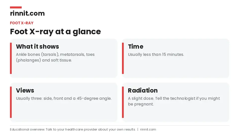
Radiation exposure is slight. Tell the technologist if you are or could be pregnant. [1]
Emergency results are often available right away; non-urgent results usually take one to two days. [1]
What is a foot X-ray?
A foot X-ray, also called a foot series or foot radiograph, creates an image of the soft tissues and bones of your foot. These include your ankle bones (tarsals), the bones at the front of your foot (metatarsals) and your toes (phalanges). [1]
How do X-rays work?
X-rays use a form of radiation called electromagnetic waves. Small beams pass through your body and form an image on special film or a digital plate. Calcium in bone absorbs more radiation, so bones look white. Soft tissues absorb less and look gray, and air looks black. X-rays are the oldest and most frequently used type of medical imaging. [1]
Why would you need a foot X-ray?
Your provider may order a foot X-ray after an injury or accident, or if you have unexplained swelling, pain or tenderness in your foot or ankle. It can help diagnose: [1]
Condition | What the X-ray may show |
Fractures | Broken bones in the foot or ankle |
Dislocations | Joints that have moved out of place |
Bunions (hallux valgus) | Joint deformity at the base of the big toe |
Arthritis | Wear and degeneration in the foot and ankle joints |
Cysts | Fluid-filled areas in bone |
Bone infection | Changes caused by infection |
Bone cancer | Bone tumors |
Sources: [1]
Foot X-rays are also used after a broken ankle has been set, to confirm the bone is lined up correctly. Bones need proper alignment to heal well. [1]

Who performs it?
A radiologic technologist (X-ray technician) performs the test. They are trained in radiation protection, patient care and positioning. [1]
How do you prepare?
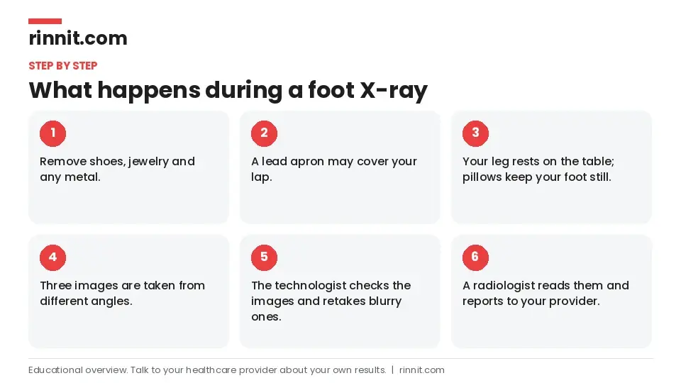
Remove clothing, shoes, jewelry or any metal that might block the image. [1]
Tell the technologist if you are pregnant or might be. Your provider will decide if the X-ray is needed and, if it is urgent, take steps to limit radiation to the baby. [1]
The technologist will explain the process and answer your questions before starting. [1]
What happens during a foot X-ray?
You go into the X-ray room, which may be in your provider's office or a hospital radiology department. The room may feel a little chilly. [1]
The technologist may place a lead apron on your lap to protect your reproductive organs. [1]
Your leg is placed on the X-ray table, and sandbags or pillows may be used to keep your foot still. Staying still prevents blurry images. [1]
The technologist steps behind a wall to take the pictures and repositions your foot between shots. [1]
Three images are usually taken: one from the side, one from the front and one at a 45-degree angle between them. Let the technologist know if you are in pain so they can help you get comfortable. The whole test usually takes less than 15 minutes. [1]
After the X-ray
You may wait a couple of minutes while the technologist checks that the images are clear. If any are blurry, they retake them right away. A radiologist, a doctor trained to read imaging, then reviews the images and sends a report to your provider. You may be referred to a foot and ankle specialist, and you may need more X-rays later to check additional views or follow your healing. [1]
What are the risks?
A foot X-ray uses only a slight amount of radiation, and side effects are rare. [1]
Pregnancy: There is a somewhat higher risk from radiation. Always tell the technologist, and a lead apron may be used. [1]
Children: They have a somewhat higher risk, so lower doses can be used. [1]
Cancer risk: Too much radiation exposure carries a small cancer risk, but providers agree the benefit of an accurate diagnosis outweighs the risk of a single X-ray. [1]
When will you get results?
Results from X-rays taken in an emergency are often available right away. For non-emergencies, the radiologist usually sends results to your provider within one to two days, and your provider then discusses them and your treatment options with you. [1]
Conclusion
A foot X-ray is a quick, simple and painless way to see what is going on inside a painful or swollen foot or ankle. It helps your provider diagnose problems such as fractures, bunions and arthritis and choose the right treatment. [1]
If you have pain after an injury, ask your provider whether you need an X-ray. Bedside checks like the Ottawa ankle rules help decide when imaging is truly needed.
Keep learning: Read our guide to the X-ray or our guide to a broken foot. For more trusted, doctor-reviewed health guides, visit Rinnit.com.
FAQ
What is a foot X-ray?
It is a quick imaging test that makes a black-and-white picture of the bones and soft tissues of your foot. [1]
What can a foot X-ray show?
It can show fractures, dislocations, bunions, arthritis, cysts, bone infections and bone tumors. [1]
Why would I need a foot X-ray?
After an injury, or for unexplained foot or ankle pain, swelling or tenderness. [1]
How do I prepare for a foot X-ray?
Remove shoes, jewelry and metal, and tell the technologist if you might be pregnant. [1]
How long does a foot X-ray take?
Usually less than 15 minutes. [1]
How many pictures are taken?
Usually three: from the side, from the front and at a 45-degree angle. [1]
Is a foot X-ray painful?
No. Tell the technologist if positioning your foot hurts so they can help. [1]
Is a foot X-ray safe?
Yes. The radiation dose is slight and side effects are rare. [1]
Can I have a foot X-ray while pregnant?
Tell the technologist first. Your provider decides if it is needed and uses precautions such as a lead apron. [1]
When will I get my foot X-ray results?
Emergency results are often immediate; non-urgent results usually take one to two days. [1]
Related Reading
Broken Foot: Symptoms, Causes, and Treatment explains foot fractures, the most common reason for a foot X-ray.
Broken Ankle: Symptoms, Causes, Treatment & Complete Guide covers ankle fractures and why follow-up X-rays check that the bone was set correctly.
Understanding Fifth Metatarsal Fractures: Types, Recovery, and Treatment explains a common break in the outer foot bone that X-rays show.
Lisfranc Injury: Understanding Midfoot Damage, Symptoms, and Recovery covers midfoot injuries that are often first checked with X-rays.
Bunions: Symptoms, Causes, and Treatment explains bunions, a joint deformity a foot X-ray can show.
Foot and Ankle Arthritis: Symptoms, Types, and Management covers the joint wear a foot X-ray can reveal.
Osteomyelitis: Symptoms, Causes, and Treatment explains bone infection, one of the conditions a foot X-ray can help find.
Bone Cancer: Symptoms, Causes, and Treatment covers bone tumors, which may first appear on an X-ray.
References
Cleveland Clinic. Foot X-Ray: Anatomy, Procedure & What to Expect. Cleveland Clinic. Last updated July 18, 2022. https://my.clevelandclinic.org/health/diagnostics/23500-foot-x-ray
Bachmann LM, Kolb E, Koller MT, et al. Accuracy of Ottawa ankle rules to exclude fractures of the ankle and mid-foot: systematic review. BMJ. 2003;326(7386):417. PMID 12595378. https://doi.org/10.1136/bmj.326.7386.417
Dowling S, Spooner CH, Liang Y, et al. Accuracy of Ottawa Ankle Rules to exclude fractures of the ankle and midfoot in children: a meta-analysis. Acad Emerg Med. 2009;16(4):277-287. PMID 19187397. https://doi.org/10.1111/j.1553-2712.2008.00333.x
Medical disclaimer
This article is for general education only and does not replace advice from a qualified healthcare professional. Talk to your doctor about your own diagnosis and treatment, and seek emergency care right away for severe or sudden symptoms.



Comments