X-Ray (Radiograph): What It Shows, How to Prepare and Is It Safe

Updated: 2 days ago
Written and medically reviewed by Dr. Baraa Alnahhal, MD · Last reviewed: November 2026 · How we review
Quick Answer: What is an X-ray?
An X-ray is a quick, painless imaging test that uses a small amount of radiation to make pictures of the inside of your body. It's used most often to check bones and joints, but it can also look at your chest, belly, spine, head and teeth. A radiologist reviews the pictures and sends a report to your provider. [1]
What the research says
Computer programs can spot a collapsed lung on chest X-rays with high accuracy. Across 23 studies covering 34,075 chest X-rays, deep learning tools detected pneumothorax (air leaking around the lung) with a pooled sensitivity of 87% and specificity of 95%. The authors see them as a way to support radiologists and speed up diagnosis. (Katzman et al., Canadian Association of Radiologists Journal, 2024) [2]
AI tools also perform well at finding pneumonia on chest X-rays. In a pooled analysis, deep learning separated pneumonia X-rays from normal ones with 98% sensitivity and 94% specificity, and told bacterial from viral pneumonia with 89% sensitivity and specificity. The authors noted study-quality concerns that need work before routine clinical use. (Li et al., Computers in Biology and Medicine, 2020) [3]
Lead shields are no longer recommended for dental X-rays. An expert committee found the dose to the reproductive organs and to a fetus from dental imaging is negligible. It advised stopping gonad, pelvic and fetal shielding, and not using thyroid collars, for standard dental X-rays and dental cone-beam CT. (Benavides et al., The Journal of the American Dental Association, 2023) [4]
Key takeaways
An X-ray uses invisible beams that pass through your body to make an image on a detector. You cannot see or feel the beams. [1]
Dense tissue like bone shows up bright white, while organs, muscle and fat show up in shades of gray. [1]
Preparation is simple: remove metal items, wear comfortable clothes or a gown, and tell your provider if you are or might be pregnant. [1]
The radiation dose is small, close to the natural background radiation you get in daily life. A dental X-ray equals about one day of background exposure. [1]
Lead aprons were used in the past, but newer evidence suggests they are not needed. [1] [4]
You can usually go back to your normal activities right after the test. [1]
What is an X-ray?
An X-ray (also called a radiograph) is a medical imaging test that uses radiation to show structures inside your body. Providers use it mainly to look at bones and joints, but it helps diagnose many other conditions too. [1]
X-rays are among the oldest reliable tools in medicine, discovered nearly 130 years ago. Today's machines make more detailed pictures and use less radiation than older ones, and X-ray technology is often combined with other tools to create more advanced images. [1]
How does an X-ray work?
During the test, a machine sends X-ray beams through the part of your body being checked. The beams land on a detector on the other side and form a picture. [1]
Different tissues absorb the beams in different amounts. Dense tissue, such as bone and some tumors, absorbs radiation easily and looks bright white. Softer tissue, such as organs, muscle and fat, appears in shades of gray. Most X-ray images are black and white. [1]

What can an X-ray show?
Providers order X-rays for many reasons, from a suspected broken bone to a stubborn cough. The body area being imaged shapes what the test can find. [1]
Body area | What it can help find |
Bone | Broken bones, dislocated joints, arthritis, bone infection and bone cancer |
Chest | Lung infections such as pneumonia and signs of congestive heart failure, often checked for chest pain, cough or shortness of breath |
Abdomen (belly) | Problems in parts of the digestive system, plus kidney and bladder stones |
Spine | Arthritis and scoliosis (a sideways curve of the spine) |
Head | Skull fractures and conditions affecting how the skull forms, such as craniosynostosis (early fusing of the skull bones) |
Teeth | Cavities and gum problems |
Sources: [1]
What are the different types of X-ray tests?
The word "X-ray" usually means a standard still picture. Several other tests also use X-rays but work in special ways. [1]
Test | How it uses X-rays |
Standard X-ray (radiograph) | Takes still pictures of a body area, most often bones, joints or the chest |
Bone density (DXA) scan | Measures bone strength and mineral content |
CT scan | Combines X-rays with a computer to build 3D images |
Dental X-ray | Checks for cavities and gum problems |
Fluoroscopy | Takes a series of X-rays to show your body moving in real time, used to diagnose problems and guide procedures such as angiograms (imaging of blood vessels) |
Mammogram | A special breast X-ray that can show changes that may point to breast cancer |
Sources: [1]
How do you prepare for an X-ray?
Most X-rays need very little preparation, though the steps can vary with the type of test. [1]
Remove metal. Take off jewelry, hairpins, hearing aids and other metal items, because metal can make the images less accurate. [1]
Dress for comfort. Wear loose, comfortable clothes, or you may be asked to change into a gown. [1]
Share your health history. Tell your provider about your medical conditions, allergies and medicines. [1]
Mention pregnancy. Let your provider know if you are pregnant or could be, so they can choose the safest test. [1]
What happens during an X-ray?
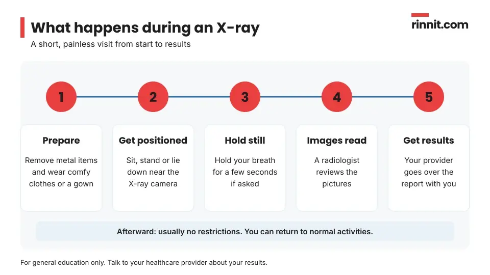
An X-ray appointment is usually short and straightforward. The exact steps depend on which body part is being imaged. [1]
You sit, stand or lie on a table, depending on the area being checked. [1]
The technologist places the X-ray camera close to that part of your body. [1]
You may be moved into a few different positions and asked to keep very still. [1]
For some images, you will hold your breath for a few seconds so the picture is not blurry. [1]
The images are taken. The beams are invisible and you will not feel anything. [1]
Young children who cannot stay still may be gently held in place with a soft restraint. This does not hurt and does not harm the child, and it lowers the chance of needing to repeat the pictures. [1]
In the past, people often wore a lead apron during X-rays. Newer evidence suggests this is not needed. [1] For dental X-rays, an expert group now advises against shielding the pelvis, reproductive organs or a fetus, and against using thyroid collars. [4]
What happens after an X-ray?
There are usually no restrictions afterward. You can go back to your normal activities right away. [1]
When will you get your X-ray results?
A radiologist reviews your images and writes a report with findings and recommendations. Your provider receives the report and goes over it with you. [1]
Timing depends on the test. Results for a bone X-ray are often available right away, though reading and recording them can take minutes to hours. More complex X-rays may take longer, and results are usually faster in an emergency. You may hear about results right after the test or see them in your electronic health record. [1]
What happens if your X-ray shows a problem?
Your next steps depend on what the X-ray shows. Your provider may recommend: [1]
Researchers are also testing computer programs that help read chest X-rays. In pooled studies, these tools found a collapsed lung and pneumonia with high accuracy, though they are meant to support, not replace, the radiologist. [2] [3]
Can an X-ray detect cancer?
An X-ray can sometimes show cancer, but it is not the main way most cancers are found or diagnosed. Tumors may be too small to see, hidden behind other structures or blended into normal tissue. [1]
Some special X-ray tests, such as mammograms, are designed to look for changes that may point to cancer in a specific area. [1]
Is an X-ray safe? Risks and radiation
X-rays are considered safe and low risk. The radiation dose is small and not much higher than the natural background radiation you are exposed to every day. A dental X-ray, for example, gives about the same amount of radiation as one day of normal background exposure. [1]

Who | What to know |
Adults | The small dose is usually safe. |
Pregnant people | Radiation can harm a developing baby, so your provider may choose another test, such as an ultrasound. |
Children | If they cannot stay still, a gentle restraint may be used. It does not hurt and helps avoid repeat images. |
Sources: [1]
When should you talk to your provider?
Ask your provider before the test if you have questions about why you need an X-ray or how it is done. [1] Speak up if any of these apply to you:
Questions to ask your provider
Why do I need this X-ray, and what are you looking for?
Do I need to do anything to prepare for this type of X-ray?
How long will the appointment take?
When and how will I get my results?
If the X-ray shows something, what tests or steps might come next?
I am pregnant or might be. Is there another test I should have instead?
Will I need a follow-up X-ray later?
Conclusion
An X-ray is one of the quickest and most familiar ways to look inside your body. It can help find broken bones, joint problems, lung infections, stones and dental issues, usually in a single short visit with little preparation. [1]
The radiation dose is small, and modern equipment uses less than in the past. Tell your provider if you are pregnant, follow their instructions and ask questions about your results so you understand what comes next. [1]
Keep learning: Read our guides to bone fractures, pneumonia and scoliosis. For more, see MedlinePlus's page on X-rays.
FAQ
What is an X-ray?
An X-ray is a painless imaging test that uses a small amount of radiation to make pictures of the inside of your body, most often your bones and joints. [1]
Why would a doctor order an X-ray?
To check for broken bones, dislocations, arthritis, lung infections, kidney or bladder stones, spine problems and dental issues. [1]
How do I prepare for an X-ray?
Remove metal items such as jewelry, wear comfortable clothes or a gown, and tell your provider about your health history and any chance of pregnancy. [1]
Does an X-ray hurt?
No. The beams are invisible and you cannot feel them. You only need to hold still, and sometimes hold your breath for a few seconds. [1]
How long does an X-ray take?
It is usually a short visit, and the exact steps depend on the body part being imaged. Bone X-ray results are often ready right away. [1]
How do I get my X-ray results?
A radiologist reads the images and sends a report to your provider, who reviews it with you. Results may also appear in your health record. [1]
Is the radiation from an X-ray dangerous?
X-rays are low risk. The dose is small, close to natural background radiation. A dental X-ray equals about one day of background exposure. [1]
Can I have an X-ray while pregnant?
Tell your provider if you are or might be pregnant. Radiation can harm a fetus, so they may choose another test, such as an ultrasound. [1]
Do I need a lead apron for an X-ray?
Lead aprons were common in the past, but newer evidence suggests they are not needed. [1]
Can an X-ray show cancer?
Sometimes, but it is not the main way most cancers are found, because tumors can be small, hidden or blend in with normal tissue. [1]
Related Reading
Learn more in our guide to Bone Fractures: What Happens When a Bone Breaks, and How It Heals.
Learn more in our guide to Pneumonia: Complete Guide to Symptoms, Causes, and Treatment.
Learn more in our guide to Scoliosis Guide: Symptoms, Causes, Diagnosis, and Treatment.
Learn more in our guide to Osteoporosis: Causes, Symptoms, Diagnosis and Treatment Explained.
References
Cleveland Clinic. X-Ray: What It Is & What It Can Show. Cleveland Clinic. Last updated January 9, 2026. https://my.clevelandclinic.org/health/diagnostics/21818-x-ray
Katzman BD, Alabousi M, Islam N, et al. Deep Learning for Pneumothorax Detection on Chest Radiograph: A Diagnostic Test Accuracy Systematic Review and Meta Analysis. Can Assoc Radiol J. 2024;75(3):525-533. PMID 38189265. https://doi.org/10.1177/08465371231220885
Li Y, Zhang Z, Dai C, et al. Accuracy of deep learning for automated detection of pneumonia using chest X-Ray images: A systematic review and meta-analysis. Comput Biol Med. 2020;123:103898. PMID 32768045. https://doi.org/10.1016/j.compbiomed.2020.103898
Benavides E, Bhula A, Gohel A, et al. Patient shielding during dentomaxillofacial radiography: Recommendations from the American Academy of Oral and Maxillofacial Radiology. J Am Dent Assoc. 2023;154(9):826-835.e2. PMID 37530694. https://doi.org/10.1016/j.adaj.2023.06.015
Medical disclaimer
This article is for general education only and does not replace advice from a qualified healthcare professional. Talk to your doctor about your own diagnosis and treatment, and seek emergency care right away for severe or sudden symptoms.



Comments