Fine-Needle Aspiration (FNA): What It Is, Why It's Done, What to Expect and Results

Updated: 2 days ago
Written and medically reviewed by Dr. Baraa Alnahhal, MD · Last reviewed: November 2026 · How we review
Quick Answer: What is fine-needle aspiration?
Fine-needle aspiration (FNA), also called a fine-needle biopsy, uses a thin needle and syringe to remove cells, tissue or fluid from a suspicious lump or abnormal area. It's the least invasive type of biopsy and is most often used for new lumps in the breast, thyroid, lymph nodes and skin. Soreness or bruising fades within a couple of days.
What the research says
Breast FNA is highly accurate when reported with a standard system. A meta-analysis of 22 studies with 10,886 people with a breast lesion found that fine-needle aspiration biopsy, reported with the IAC Yokohama System, had an overall accuracy (area under the curve) of 0.975, with specificity of 98.3% when suspicious or malignant results were counted as positive. (Paul et al., Acta Cytologica, 2023) [2]
Indeterminate thyroid FNA results carry a real but variable cancer risk. A meta-analysis of 38 studies covering 145,066 thyroid fine-needle aspirations found the risk of cancer in indeterminate nodules was 25.4% in Western series and 41.9% in Asian series, reflecting different follow-up and surgery practices. (Vuong et al., Cancer Cytopathology, 2020) [3]
Key takeaways
Fine-needle aspiration uses a thin needle and syringe to sample cells, tissue or fluid from a lump [1].
It is the least invasive type of biopsy, but it has limits [1].
It is most often used for new lumps in the breast, thyroid, lymph nodes and skin [1].
With ultrasound, CT or endoscopic ultrasound guidance, it can sample the pancreas, lungs and other deep areas [1].
FNA can also treat cysts, abscesses and seromas by draining fluid [1].
Soreness and bruising are common and minor; bleeding, infection and damage to nearby structures are rare [1].
What is fine-needle aspiration?
Fine-needle aspiration (FNA) uses a thin needle and a syringe to collect a sample of cells, tissue or fluid from a suspicious lump or abnormal area [1]. It is also called a fine-needle biopsy [1].
The sample goes to a cytology lab, where a cytotechnologist screens the cells and a pathologist examines them [1]. FNA is the least invasive type of biopsy, but it has limits [1].
FNA vs core needle biopsy

Feature | Fine-needle aspiration | Core needle biopsy |
Needle | A thin needle [1] | A wider needle, sometimes spring-loaded [1] |
Sample size | Cells, tissue or fluid [1] | More tissue, which can give more information [1] |
Incision | None [1] | Sometimes a small incision [1] |
Common use | Many areas of the body [1] | Most often breast biopsies [1] |
Sources: [1]
Why fine-needle aspiration is done
FNA has two main purposes: diagnosing a problem and treating one [1]. It can also help guide treatment when cancer has spread, because pathologists can look for genetic or molecular markers that predict how a cancer will respond to certain treatments [1].
Diagnostic FNA
FNA can be used whenever a tissue or fluid sample would help make a diagnosis [1]. It can help diagnose [1]:
It is most often used on newly found lumps in the breast, including breast cysts, the thyroid, including thyroid nodules, lymph nodes and skin [1]. With imaging guidance such as endoscopic ultrasound or a CT scan, providers can also sample deeper areas, including the pancreas, digestive tract, esophagus, windpipe, lungs and mediastinum, the space in the chest that holds the heart [1].
Therapeutic FNA
FNA can treat some lumps by draining fluid from them, such as [1]:
Providers may choose FNA over a standard cut-and-drain procedure in cosmetically sensitive areas such as the breast [1]. Because FNA can spread infection, it cannot be used in every case [1].
How to prepare for fine-needle aspiration
You usually do not need any special preparation [1]. Your provider will explain what to expect and how to get ready [1]. Bleeding risk is highest in people with clotting disorders or those taking blood thinners, so mention these to your provider [1].
What happens during fine-needle aspiration
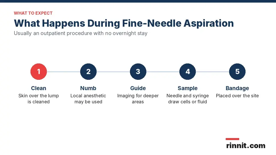
FNA is usually an outpatient procedure done in a provider's office or hospital, with no overnight stay [1]. The exact steps depend on the area, but generally [1]:
You lie or sit on an exam table and the skin is cleaned [1].
A local anesthetic may be injected to numb the area. You may still feel a pinch or some discomfort [1].
For deeper areas, ultrasound, CT or a mammogram guides the needle [1].
The needle is inserted and the syringe draws out cells, tissue or fluid [1].
Several samples may be taken, with a new needle and syringe each time [1].
A bandage is placed over the site [1].
Risks of fine-needle aspiration
Soreness and bruising at the site are common, usually minor and gone within a couple of days [1]. Serious complications are relatively rare and can include [1]:
Fine-needle aspiration results
A pathologist studies the sample under a microscope and may run other tests, then sends a report to your provider [1]. Possible results include [1]:

Sometimes the lab asks for more samples to give a more accurate report [1]. Your provider will explain the result and plan next steps with you [1].
When to call your provider
Contact your provider if you notice signs of infection, such as fever, pus or redness at the site, or if the lump changes in an unexpected way after the FNA [1].
Conclusion
Fine-needle aspiration is a quick, minimally invasive way to find out what a lump is, and in some cases to drain it at the same time. It is most often used for lumps in the breast, thyroid, lymph nodes and skin.
Side effects are usually limited to mild soreness or bruising. Waiting for results can be stressful, so ask your provider when to expect them and what the possible next steps are.
Keep learning: Read our overview of the biopsy, our guide to the thyroid ultrasound or our guide to the breast biopsy. For more background, see the RadiologyInfo general biopsy guide.
FAQ
What is fine-needle aspiration?
It uses a thin needle and syringe to remove cells, tissue or fluid from a lump so a pathologist can examine it.
Is fine-needle aspiration the same as a biopsy?
Yes. It is a type of biopsy, also called a fine-needle biopsy, and the least invasive kind.
What is the difference between FNA and a core biopsy?
A core biopsy uses a wider needle and takes more tissue. FNA uses a thin needle and is used in many body areas.
What is FNA used for?
To diagnose cancer, infections and noncancerous lumps, and to drain cysts, abscesses and seromas.
Which areas are commonly tested with FNA?
New lumps in the breast, thyroid, lymph nodes and skin. Deeper areas can be reached with imaging guidance.
Does fine-needle aspiration hurt?
A numbing medicine may be used. You may feel a pinch or discomfort, and the site may be sore for a couple of days.
Do I need to prepare for FNA?
Usually not. Tell your provider if you have a bleeding disorder or take blood thinners.
What are the risks of fine-needle aspiration?
Soreness and bruising are common. Bleeding, infection, damage to nearby structures and fistula are rare.
What can FNA results show?
No abnormal cells, a mix of normal and abnormal cells, precancerous or cancer cells, or signs of infection.
When should I call my doctor after FNA?
Call for fever, pus or redness at the site, or if the lump changes unexpectedly.
Related Reading
Thyroid Nodules: Symptoms, Causes, Diagnosis and Treatment — What You Need to Know explains thyroid nodules, one of the most common lumps tested with fine-needle aspiration.
Breast Cysts: Symptoms, Causes, and Treatment covers breast cysts, which FNA can both diagnose and drain.
Swollen Lymph Nodes: Symptoms, Causes, and When to Seek Care explains swollen lymph nodes, a common reason to sample a node with a thin needle.
Lipoma: Symptoms, Causes, Diagnosis, and Treatment covers lipomas, a noncancerous fatty lump FNA can help identify.
Bone & Joint Infections (Septic Arthritis): Symptoms, Causes, Diagnosis & Treatment — Complete Guide explains septic arthritis, a joint infection a needle sample can help diagnose.
Thyroid Cancer: Symptoms, Causes, Diagnosis and Treatment — What You Need to Know covers thyroid cancer, which is often first diagnosed with fine-needle aspiration.
References
Cleveland Clinic. Fine-Needle Aspiration (FNA): What It Is, Procedure & Results. Cleveland Clinic. Last updated May 16, 2023. https://my.clevelandclinic.org/health/diagnostics/17872-fine-needle-aspiration-fna
Paul P, Azad S, Agrawal S, et al. Systematic Review and Meta-Analysis of the Diagnostic Accuracy of the International Academy of Cytology Yokohama System for Reporting Breast Fine-Needle Aspiration Biopsy in Diagnosing Breast Cancer. Acta Cytol. 2023;67(1):1-16. PMID 36412573. https://doi.org/10.1159/000527346
Vuong HG, Ngo HTT, Bychkov A, et al. Differences in surgical resection rate and risk of malignancy in thyroid cytopathology practice between Western and Asian countries: A systematic review and meta-analysis. Cancer Cytopathol. 2020;128(4):238-249. PMID 31883438. https://doi.org/10.1002/cncy.22228
Medical disclaimer
This article is for general education only and does not replace advice from a qualified healthcare professional. Talk to your doctor about your own diagnosis and treatment, and seek emergency care right away for severe or sudden symptoms.



Comments