Chest X-Ray (CXR): What It Shows, How It's Done and What Results Mean

Written and medically reviewed by Dr. Baraa Alnahhal, MD · Last reviewed: November 2026 · How we review
Quick Answer: What is a chest X-ray?
A chest X-ray, also called a chest radiograph or CXR, is a quick, noninvasive imaging test that makes pictures of your heart, lungs and bones. It helps providers find problems such as pneumonia, emphysema, COPD, heart failure and broken ribs. The test takes a few minutes, and nonemergency results usually come back in one to two days. [1]
What the research says
A chest X-ray finds most, but not all, tuberculosis in community screening. In a reanalysis of 21 tuberculosis screening studies, a chest X-ray picked up 86% of confirmed cases among people with symptoms, lower than the 94% reported in the original analysis. The authors say the real-world figure for screening everyone is likely lower still. (Burua et al., The International Journal of Tuberculosis and Lung Disease, 2026) [2]
A chest X-ray taken lying down can miss a collapsed lung. In a review of eight studies with 1,048 adults suspected of having a pneumothorax, a chest X-ray taken lying on the back found 50.2% of cases, while bedside ultrasound found 90.9%. The X-ray rarely flagged a pneumothorax that was not there (99.4% specificity). (Alrajhi et al., Chest, 2012) [3]
Key takeaways
A chest X-ray is a quick, noninvasive test that makes images of your heart, lungs and bones. [1]
It is often one of the first tests used for chest pain, a lasting cough, trouble breathing or a possible broken rib. [1]
You need little to no preparation; just wear loose clothing without metal and leave jewelry at home. [1]
Most chest X-rays include two images, one from the front and one from the side, and you hold your breath for each. [1]
The test uses a very small amount of radiation, but tell your provider if there is any chance you are pregnant. [1]
An abnormal result may lead to more tests, such as a CT scan or PET scan. [1]
What is a chest X-ray?
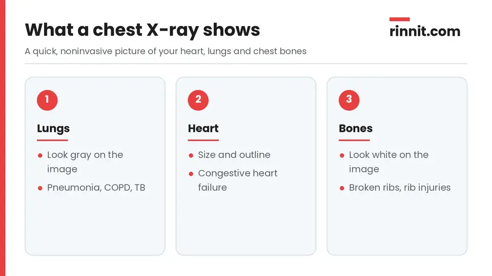
A chest X-ray shows your heart, lungs and the bones of your chest.
A chest X-ray is an imaging test that creates pictures of your heart, lungs and bones. You may also hear it called a chest radiograph or CXR. [1]
Different tissues in your body absorb radiation in different amounts. On the image, bones look white and lungs look gray, and providers read these colors and shades to look for problems. [1]
Why would you need a chest X-ray?
Your provider may order a chest X-ray if you have symptoms such as: [1]
Chest pain
A chronic (long-lasting) cough
Difficulty breathing (dyspnea)
Fever along with other signs of infection
A chest injury, such as a possible broken rib
A chest X-ray can help diagnose or check on conditions such as: [1]
Area of the chest | Conditions a chest X-ray can help find |
Lungs | Pneumonia, emphysema or COPD, lung cancer and tuberculosis (TB) |
Heart | Congestive heart failure |
Bones | Rib cage injuries, such as broken ribs |
Sources: [1]
How do you prepare for a chest X-ray?
A chest X-ray needs little to no preparation. To get ready: [1]
Wear loose clothing without metal, such as zippers, snaps or bra closures.
Leave jewelry at home.
Ask the X-ray center whether you need to remove any body piercings.
Tell your provider if you are pregnant or think you might be, even slightly.
What happens during a chest X-ray?
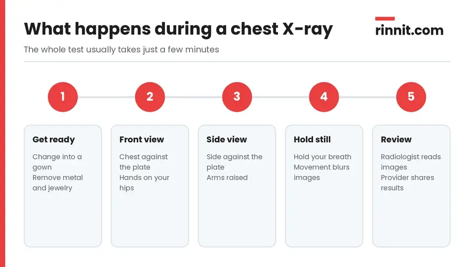
The test usually takes just a few minutes.
A radiology technologist performs the test. You may change into a gown and remove any metal around your chest, such as a necklace. [1]
Most chest X-rays include two views: [1]
View | How you are positioned |
Front view | Your chest presses against the imaging plate, with your hands on your hips |
Side view | Your side presses against the plate, with your arms raised |
Sources: [1]
For each image, you stay very still and hold your breath, because any movement can blur the picture. The whole test takes a few minutes. [1]
Sometimes a provider orders four images, adding views taken while you lie on your back and on your side. [1]
After the images are taken, the technologist may check them and retake any that are blurry. A radiologist then reviews the images, and your provider goes over the images and report with you. [1]
Is a chest X-ray safe?
A chest X-ray uses a very small amount of radiation. Providers use only the amount needed to get clear, good-quality images. [1]
Radiation can harm a developing fetus, so tell your provider if you might be pregnant. A simple chest X-ray uses so little radiation that it usually causes no problems, but providers prefer to limit radiation until after delivery and will decide based on your symptoms. [1]
What do chest X-ray results mean?
In a nonemergency, results usually take one to two days. In an emergency, you often get results within a few minutes or hours. [1]
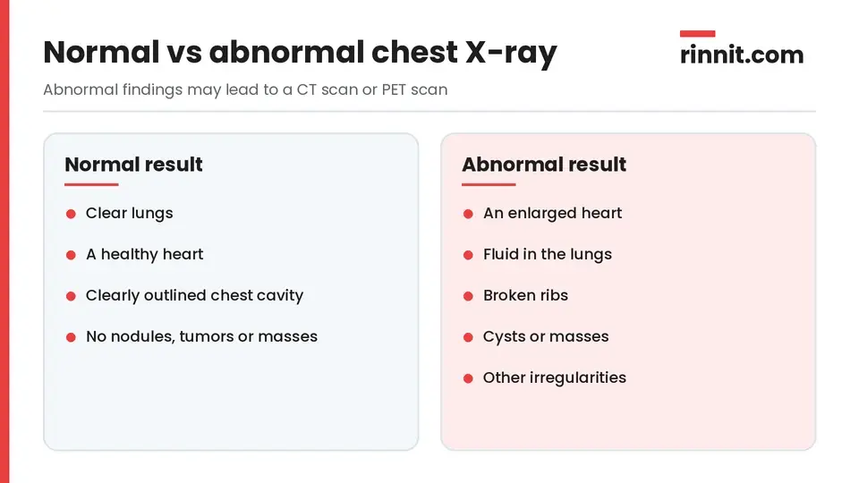
A radiologist compares your images with what a healthy chest looks like.
Result | What it may show |
Normal | Clear lungs, a healthy heart, a clearly outlined chest cavity and no visible nodules, tumors or masses |
Abnormal | An enlarged heart, fluid in the lungs, broken ribs, cysts, masses or other irregularities |
Sources: [1]
What happens after an abnormal result?
An abnormal chest X-ray may lead to more tests, such as a CT scan or a PET scan, to look more closely at what was found. Your provider will use the results to help decide on treatment. [1]
When should you call your provider?
Contact your provider if you have a chest injury such as a possible broken rib, chest pain that does not go away, a chronic cough or difficulty breathing. [1]
Conclusion
A chest X-ray is a fast, noninvasive way to check your lungs, heart and rib cage. It is often one of the first tests providers use for broken bones and for lung and heart problems.
If your result is abnormal, ask your provider what it means and whether you need a follow-up test such as a CT scan.
Keep learning: Read our guide to pneumonia or our overview of X-rays in general. For more trusted, doctor-reviewed health guides, visit Rinnit.com.
FAQ
What is a chest X-ray?
It is a quick, noninvasive imaging test that makes pictures of your heart, lungs and bones.
What can a chest X-ray detect?
It can help find pneumonia, emphysema or COPD, heart failure, lung cancer, tuberculosis and rib injuries.
How do I prepare for a chest X-ray?
Little preparation is needed; wear loose clothing without metal and leave jewelry at home.
How long does a chest X-ray take?
The test itself takes only a few minutes.
How many images are taken?
Usually two, one from the front and one from the side, though sometimes four are taken.
Why do I need to hold my breath?
Holding still and holding your breath keeps the images from blurring.
Is a chest X-ray safe?
Yes, it uses a very small amount of radiation, only as much as needed for clear images.
Can I have a chest X-ray if I am pregnant?
Tell your provider first; the dose is very low, but providers prefer to limit radiation during pregnancy.
How long do chest X-ray results take?
Usually one to two days, or within minutes to hours in an emergency.
What happens if my chest X-ray is abnormal?
Your provider may order more tests, such as a CT scan or PET scan.
Related Reading
Pneumonia: Complete Guide to Symptoms, Causes, and Treatment explains the lung infection that a chest X-ray helps providers diagnose.
What Is Chronic Obstructive Pulmonary Disease (COPD)? Symptoms, Causes, Stages, and Treatment covers COPD and emphysema, two conditions a chest X-ray can help diagnose.
Congestive Heart Failure: Symptoms, Stages, and How It's Treated describes a heart condition that a chest X-ray can help diagnose.
Lung Cancer: Symptoms, Causes, Diagnosis, and Treatment Explained explains lung cancer, which can show up as a mass on a chest X-ray.
Tuberculosis (TB): Symptoms, Causes, Testing, Treatment and Prevention covers the lung infection that chest X-rays help detect and screen for.
Rib Fracture: Symptoms, Causes, and Recovery Guide explains broken ribs, a common reason for a chest X-ray after an injury.
Enlarged Heart: Symptoms, Causes, and Treatment describes a finding that an abnormal chest X-ray can reveal.
Sniff Test: Diaphragm Fluoroscopy for Weakness and Paralysis explains the X-ray test that checks how the diaphragm moves.
References
Cleveland Clinic. Chest X-Ray (CXR): What It Is, What To Expect & Results. Cleveland Clinic. Last updated November 20, 2023. https://my.clevelandclinic.org/health/diagnostics/10228-chest-x-ray
Burua A, Lukoye D, Kirenga B, et al. Sensitivity of chest X-ray from community screening: a meta-reanalysis of TB prevalence survey data. Int J Tuberc Lung Dis. 2026;30(9):396-402. PMID 42665896. https://doi.org/10.5588/ijtld.25.0744
Alrajhi K, Woo MY, Vaillancourt C. Test characteristics of ultrasonography for the detection of pneumothorax: a systematic review and meta-analysis. Chest. 2012;141(3):703-708. PMID 21868468. https://doi.org/10.1378/chest.11-0131
Medical disclaimer
This article is for general education only and does not replace advice from a qualified healthcare professional. Talk to your doctor about your own diagnosis and treatment, and seek emergency care right away for severe or sudden symptoms.



Comments