Brain MRI: Why It's Done, How to Prepare and What to Expect

Updated: 2 days ago
Written and medically reviewed by Dr. Baraa Alnahhal, MD · Last reviewed: November 2026 · How we review
Quick Answer: What is a brain MRI?
A brain MRI (magnetic resonance imaging) is a painless scan that makes clear pictures of the structures inside the head, mainly the brain. It uses a large magnet, radio waves and a computer instead of radiation. Providers use it to diagnose and monitor stroke, brain tumors, multiple sclerosis and brain aneurysms. It takes about 30 to 60 minutes. [1]
What the research says
MRI finds acute stroke far more often than CT. In 356 patients assessed in the emergency department for suspected stroke, MRI detected acute ischemic stroke in 46% versus 10% with CT, and its sensitivity for any acute stroke was 83% versus 26% for CT. (Chalela et al., The Lancet, 2007) [2]
Brain MRIs often turn up unexpected findings. Among 2,000 adults from the general population (average age 63), MRI showed silent brain infarcts in 7.2%, cerebral aneurysms in 1.8% and benign tumors, mostly meningiomas, in 1.6%. (Vernooij et al., The New England Journal of Medicine, 2007) [3]
Key takeaways
A brain MRI is the most sensitive imaging test for the brain compared with CT scans or X-rays. [1]
It doesn't use radiation; it relies on a strong magnet and radio waves. [1]
Some scans use contrast, often gadolinium, injected through an IV to sharpen the images. [1]
Tell your team about pacemakers, cochlear implants, aneurysm clips, metal fragments, pregnancy or claustrophobia before the scan. [1]
The scan takes 30 to 60 minutes, and the report is usually ready within one or two days. [1]
What is a brain MRI?
A brain MRI is a painless procedure that produces very clear images of the structures inside your head. It uses a large magnet, radio waves and a computer, and it doesn't use radiation. MRI is currently the most sensitive imaging test of the head, especially the brain, compared with CT scans and X-rays. [1]
A head MRI and a brain MRI are the same procedure. Both show the inside of your head, including your brain, facial bones, blood vessels and nerves. For how MRI works on other parts of the body, see our MRI guide. [1]
What is a brain MRI with contrast?
Some brain MRIs use an injection of contrast material, often gadolinium, a rare earth metal. It changes the magnetic properties of nearby water molecules and makes the images clearer. Contrast improves the view of tumors, inflammation, blood vessels and the blood supply of certain organs, and helps diagnose multiple sclerosis, stroke, dementia and infection. [1]
Contrast is given through an IV in your hand or arm. Contrast materials are considered safe; side effects can range from mild to severe, but severe reactions are very rare. [1]
What does a brain MRI show?

A brain MRI shows far more than the brain itself.
A brain MRI shows the structures inside your head, including: [1]
Your brain and the blood vessels that connect to it
Your skull and facial bones
Structures in your inner ear
Your eyes and their supporting tissues, such as your optic nerves
Large nerves in your head (cranial nerves)
Surrounding soft tissues such as fat, muscle and connective tissue
It can reveal inflammation and swelling, structural problems, abnormal growths or masses, fluid leaks, bleeding in the brain and white matter disease. [1]
Why would you need a brain MRI?
Neurologists and other providers order brain MRIs to diagnose new conditions or monitor existing ones, including: [1]
Stroke, a blood clot in the brain or brain bleeding (hemorrhage)
Brain aneurysm
Brain tumors and cysts
Multiple sclerosis (MS) and other chronic neurological conditions
Brain damage linked to epilepsy
Brain infections such as encephalitis
Dementia
Hydrocephalus
Pituitary gland problems, such as a pituitary adenoma
Problems with brain development or structure, such as Chiari malformation
Traumatic brain injury (TBI)
Your provider may also order one for migraines or chronic headaches, seizures, vertigo or frequent severe dizziness, unexplained hearing loss, vision problems not explained by an eye exam, hormonal imbalances related to the hypothalamus or pituitary gland, big changes in thinking or behavior, or extreme weakness and fatigue. Brain MRIs are also used before and after head surgery and after significant head injuries. [1]
How do you prepare for a brain MRI?
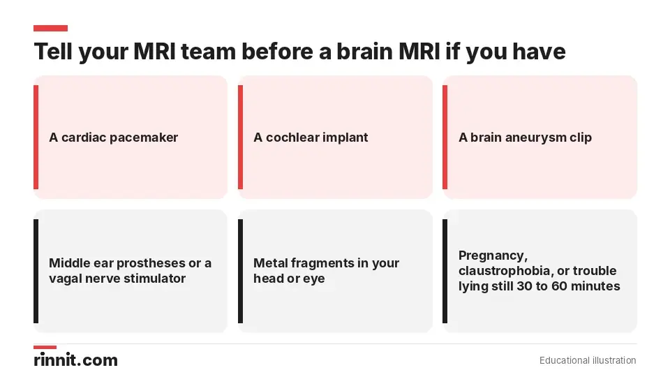
The MRI magnet can affect metal and implanted devices, so safety screening comes first.
Rules about eating and drinking vary by the reason for your scan. Eat and take your medicines as usual unless your provider tells you otherwise. [1]
The scanner's strong magnet and radio waves can heat or move some metal objects and can make some implanted electronic devices malfunction. Tell your provider and MRI technologist if you have: [1]
A cardiac pacemaker
Middle ear prostheses or a cochlear implant
A clip used for a brain aneurysm
A vagal nerve stimulator
Metal fragments in your head or eyeball
Also tell them if you're pregnant, can't lie on your back for 30 to 60 minutes or have claustrophobia (fear of enclosed spaces). Some implants need special scheduling or an X-ray first to check their exact location. [1]
What should you leave at home?
Remove or leave behind metal and electronic items, which can be damaged, distort the images, cause burns or become dangerous projectiles: [1]
Jewelry, watches, credit cards and hearing aids
Pins, metal hair accessories, underwire bras and metal zippers
Removable dental work such as dentures
Pens, pocketknives and eyeglasses
Body piercings
Cell phones, electronic watches and tracking devices
What happens during a brain MRI?
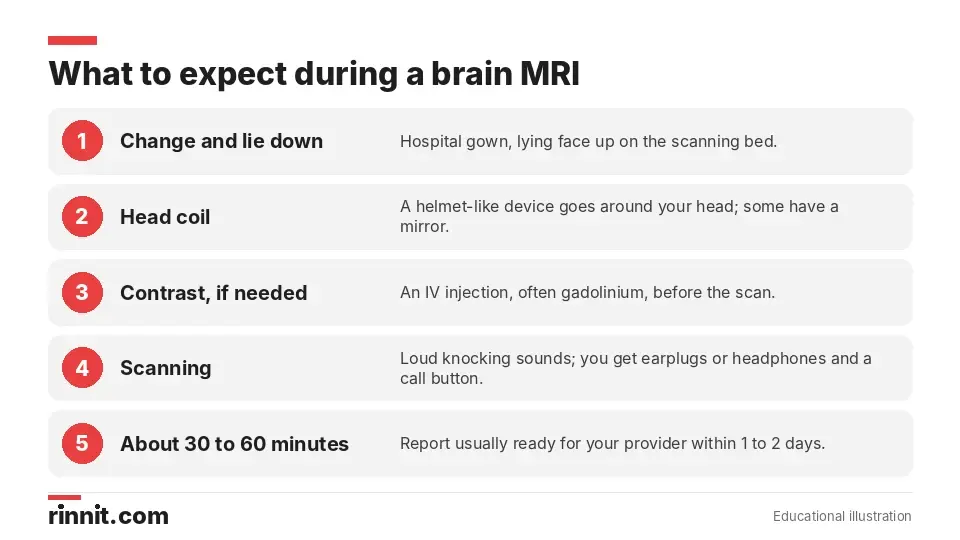
The main challenges are staying still and the noise; the scan itself is painless.
A radiologist or radiology technologist performs the scan. Here's what to expect: [1]
You change into a hospital gown and lie face up on the scanning bed.
The technologist places a helmet-like head coil around your head. Some have a mirror or small screen, which can help with claustrophobia.
The bed slides into the MRI machine. For a brain MRI, your whole body usually doesn't go into the tunnel.
You hear loud knocking and clicking sounds for several minutes at a time. You'll wear earplugs or headphones, and may be able to listen to music.
You stay very still. The area being scanned may feel slightly warm.
The technologist can see and talk with you the whole time through an intercom, and you'll hold a call button. If your scan needs contrast, you'll get an IV injection; some people notice a brief metallic taste. If you have claustrophobia, your provider may offer a sedative or, in some cases, anesthesia. [1]
How long does a brain MRI take?
About 30 minutes to an hour. It may take longer if contrast is used. [1]
Results and follow-up
A radiologist analyzes the images and sends a signed report to your provider, usually within one or two days. Your provider will share the results and explain whether you need a follow-up exam. [1]
Because MRI is so detailed, it sometimes finds things nobody was looking for, such as silent old strokes or small aneurysms. Ask your provider whether any finding needs action or simply monitoring. [3]
Conclusion
A brain MRI is a safe, painless and highly detailed way to look inside your head without radiation. It helps diagnose stroke, tumors, MS, aneurysms and many other conditions, and to track how they change over time. [1] [2]
Good preparation makes the scan go smoothly: share your full implant and metal history, leave metal at home and tell your team if you're anxious about enclosed spaces. [1]
Keep learning: Read our stroke guide or our multiple sclerosis guide. For more background, see RadiologyInfo's head MRI page.
FAQ
What is a brain MRI?
A painless scan that uses a strong magnet, radio waves and a computer to make detailed pictures of your brain and other structures in your head. [1]
Is a brain MRI the same as a head MRI?
Yes. They are the same procedure. [1]
Does a brain MRI use radiation?
No. It uses a magnet and radio waves, not radiation. [1]
What can a brain MRI detect?
Stroke, bleeding, aneurysms, tumors, multiple sclerosis, infections, hydrocephalus, dementia, injuries and structural problems. [1]
How long does a brain MRI take?
About 30 minutes to an hour, or longer with contrast. [1]
Why is contrast used for a brain MRI?
Contrast, often gadolinium, makes tumors, inflammation and blood vessels easier to see. [1]
Can I eat before a brain MRI?
Usually yes. Eat and take your medicines as normal unless your provider tells you otherwise. [1]
Who should not have a brain MRI?
People with certain implants, such as some pacemakers, cochlear implants or aneurysm clips, need special review first. [1]
What if I'm claustrophobic?
Tell your provider. Head coils with mirrors or screens can help, and a sedative or anesthesia may be offered. [1]
When will I get my brain MRI results?
The radiologist's report usually reaches your provider within one or two days. [1]
Related Reading
Stroke: Symptoms, Causes, Diagnosis, and Treatment — A Complete Guide explains the emergency that brain MRI detects better than CT.
Brain Aneurysm: Symptoms, Causes, and Treatment covers a bulging blood vessel that MRI can find.
Brain Tumor: Symptoms, Causes, Diagnosis and Treatment — What You Need to Know explains growths that brain MRI helps diagnose and monitor.
What Is Multiple Sclerosis? A Plain-Language Guide to Symptoms, Types, and How It's Treated covers a condition diagnosed and tracked with MRI.
Epilepsy: Seizures, Symptoms, Causes, and Treatment explains seizures that may lead to a brain MRI.
Hydrocephalus: Symptoms, Causes & Treatment — What You Need to Know covers fluid buildup in the brain that MRI can show.
Chiari Malformation: Types, Symptoms, Causes, Diagnosis & Treatment Guide explains a structural brain problem diagnosed with MRI.
Traumatic Brain Injury: Symptoms, Emergency Signs, Diagnosis and Recovery covers head injuries that may need a brain MRI.
Neurological Exam: What It Checks, What to Expect and What Happens Next explains the hands-on exam of the brain, spinal cord and nerves.
Gadolinium Contrast for MRI: How It Works, Side Effects and Kidney Safety explains the MRI contrast dye, its side effects and kidney safety.
References
Cleveland Clinic. Brain MRI: What It Is, Purpose, Procedure & Results. Cleveland Clinic. Last updated May 9, 2022. https://my.clevelandclinic.org/health/diagnostics/22966-brain-mri
Chalela JA, Kidwell CS, Nentwich LM, et al. Magnetic resonance imaging and computed tomography in emergency assessment of patients with suspected acute stroke: a prospective comparison. Lancet. 2007;369(9558):293-298. PMID 17258669. https://doi.org/10.1016/S0140-6736(07)60151-2
Vernooij MW, Ikram MA, Tanghe HL, et al. Incidental findings on brain MRI in the general population. N Engl J Med. 2007;357(18):1821-1828. PMID 17978290. https://doi.org/10.1056/NEJMoa070972
Medical disclaimer
This article is for general education only and does not replace advice from a qualified healthcare professional. Talk to your doctor about your own diagnosis and treatment, and seek emergency care right away for severe or sudden symptoms.



Comments