Visual Field Test: Types, What to Expect and What Results Mean

Updated: 2 days ago
Written and medically reviewed by Dr. Baraa Alnahhal, MD · Last reviewed: November 2026 · How we review
Quick Answer: What is a visual field test?
A visual field test, also called perimetry, is a simple, painless test that measures peripheral (side) vision. You look straight ahead at a fixed point and signal when you see a light or object off to the side. It can find glaucoma early, before you notice any vision loss. It takes 5 to 10 minutes per eye. [1]
What the research says
Regular repeat testing is needed to spot glaucoma getting worse. Expert recommendations based on statistical modeling found that about three visual field tests per year are needed to detect a 4-decibel loss in mean deviation over 2 years in a person with average test variability. (Chauhan et al., British Journal of Ophthalmology, 2008) [2]
Test-to-test variability can delay detection of progression. In 47,003 automated perimetry tests from 5,402 eyes, results varied more from test to test in Black (1.80 dB) and Hispanic (1.81 dB) participants than in White (1.56 dB) or non-Hispanic (1.52 dB) participants, and higher variability delayed detection of glaucoma progression. (Beniz et al., Scientific Reports, 2024) [3]
Key takeaways
A visual field test measures how far you can see to the sides while looking straight ahead, and how sensitive your vision is across that area. [1]
It is a key test for finding and tracking glaucoma, which often causes no symptoms early on. [1]
Common types include the Amsler grid, confrontation test, kinetic perimetry and automated static perimetry. [1]
No preparation is needed, and the test usually takes 5 to 10 minutes per eye. [1]
Go to the emergency room right away for sudden vision loss or eye pain. [1]
What is a visual field test?
A visual field test measures your peripheral vision in two ways: how far up, down, left and right you can see while looking straight ahead, and how sensitive your vision is across your whole visual field. [1]
Vision is usually sharpest in the center. Objects get harder to see the farther they move from the center and disappear once they move far enough to the side. [1]
Why is a visual field test done?
It is part of a routine eye exam with an optometrist or ophthalmologist. It can detect glaucoma early, because many people with glaucoma notice no symptoms while their side vision is already getting worse. [1]
The test also helps check your retina, optic nerve and brain, which work together to turn light into the images you see. Known patterns of vision loss help identify certain injuries or diseases. [1]
Conditions a visual field test can help evaluate
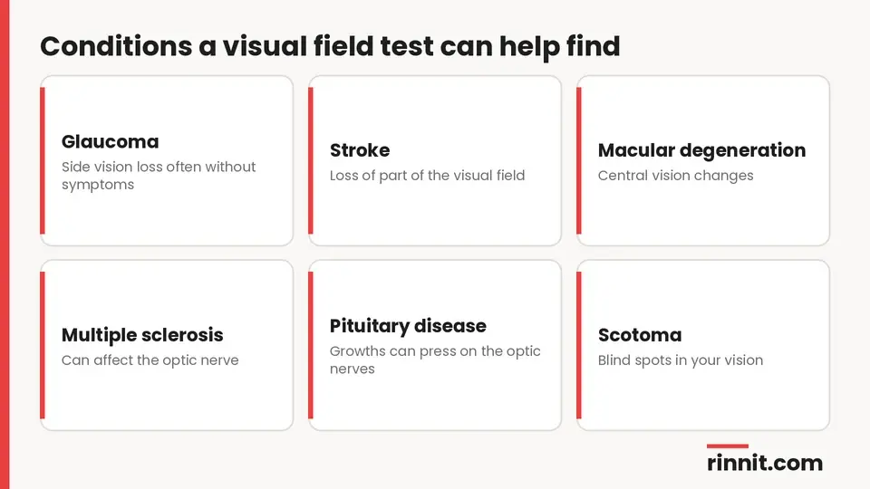
Glaucoma
Stroke
Macular degeneration
Multiple sclerosis (MS)
Graves' disease
Pituitary gland disease
Blind spots (scotoma)
Sources: [1]
What are the types of visual field tests?

Test | How it works | What it checks |
Amsler grid | You stare at a central dot on a grid of straight lines and report any blurry, wavy or broken areas | Central vision only |
Confrontation test | The examiner sits about 3 to 4 feet (around 1 meter) away and moves their hands inward from the sides | The outer edge of the visual field; not very precise |
Kinetic perimetry | An object is moved through your visual field and you signal when you see it (often the Goldmann test) | The boundaries of your visual field |
Static (automated) perimetry | You look into a bowl-shaped machine and press a button when you see a light (such as Humphrey or Octopus) | Sensitivity across the whole field, compared with normal standards |
Sources: [1]
Computerized machines give more accurate and repeatable results because your head stays in a fixed position, a central target steadies your gaze, tiny lights of adjustable brightness and color are used, and each result is compared with established normal values. [1]
How do you prepare for a visual field test?
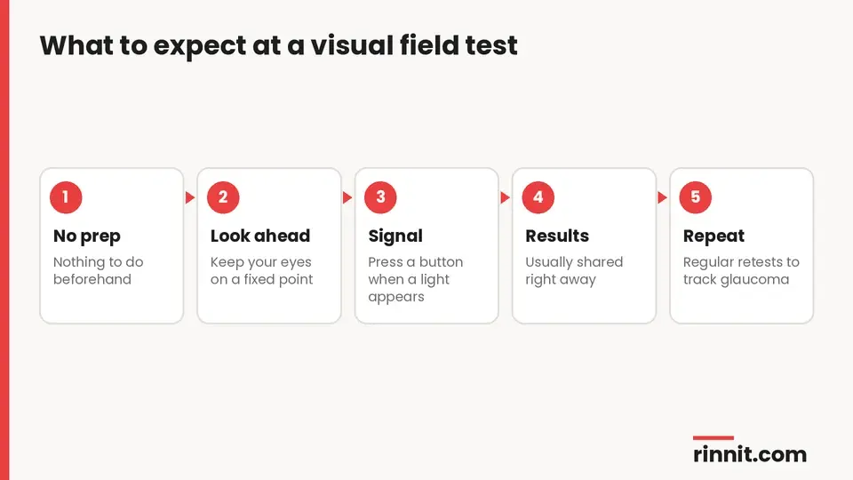
No preparation is needed. Your provider will explain where to look and how to respond. [1]
What happens during a visual field test?
You look straight ahead at a fixed point and signal when you see a light or object appear off to the side. With automated perimetry, you press a button each time you see a light. The test usually takes no more than about 5 to 10 minutes per eye. [1]
Are there any risks?
No. The test is noninvasive, so side effects are unlikely. [1]
What do visual field test results mean?
Result | What it means |
Normal | Your side vision is about as good as that of people without vision problems |
Abnormal | Patterns of dots or numbers show where and how much vision is lost, from a small area to large areas |
Sources: [1]
Providers usually share results right away. An abnormal result may point to conditions such as glaucoma, macular degeneration or stroke, and your provider will discuss next steps and treatment. [1]
Why might you need to repeat the test?
Your provider may repeat the test right away if results seem unreliable, for example if you were tired. They may also repeat it after a few weeks, months or a year, and regularly during treatment to track changes. This is especially important for glaucoma, so keep all scheduled appointments. [1]
When should you contact your eye care provider?
Report any new vision loss or eye discomfort to your eye care provider. If you have sudden vision loss or eye pain, go to an emergency room right away. [1]
Conclusion
A visual field test is a quick, painless way to map your side vision. Because glaucoma can steal peripheral vision without symptoms, this test is one of the best ways to catch it early and track it over time.
Keep up with regular eye exams and repeat testing if your provider recommends it, and seek urgent care for any sudden change in your vision.
Keep learning: Read our guides to glaucoma tests, the eye exam and tonometry. For more background, see the MedlinePlus page on the visual field test.
FAQ
What is a visual field test?
A painless test that measures your peripheral (side) vision while you look straight ahead. [1]
What does a visual field test detect?
It can help detect glaucoma, stroke, macular degeneration, MS, Graves' disease, pituitary disease and blind spots. [1]
How long does a visual field test take?
Usually no more than about 5 to 10 minutes per eye. [1]
Do I need to prepare for a visual field test?
No preparation is needed. [1]
Is a visual field test painful?
No; it is noninvasive and side effects are unlikely. [1]
What is the Humphrey visual field test?
An automated static perimetry test where you look into a bowl-shaped machine and press a button when you see a light. [1]
What does an abnormal visual field test mean?
It shows where and how much side vision is lost and may point to glaucoma, macular degeneration or stroke. [1]
Why do I need to repeat my visual field test?
To confirm unreliable results and to track changes over time, especially with glaucoma. [1]
How soon will I get my results?
Providers usually share results right away. [1]
When should I seek urgent care for vision changes?
Go to the emergency room for sudden vision loss or eye pain. [1]
Related Reading
Glaucoma: The Silent Threat to Vision, and How Early Treatment Can Protect Your Sight explains the main condition visual field testing tracks.
What Is Open-Angle Glaucoma? Symptoms, Causes, and Treatment covers the most common glaucoma type, which slowly narrows side vision.
Scotoma: Understanding Blind Spots in Your Vision explains the blind spots a visual field test can map.
Dry Macular Degeneration: Complete Guide to Symptoms, Causes & Treatment covers central vision loss that an Amsler grid can show.
Stroke: Symptoms, Causes, Diagnosis, and Treatment — A Complete Guide explains how a stroke can cut off part of your visual field.
Pituitary Tumors and Adenomas: Types, Symptoms, and Treatment Guide covers pituitary growths that can press on the optic nerves.
What Is Multiple Sclerosis? A Plain-Language Guide to Symptoms, Types, and How It's Treated explains a condition that can affect the optic nerve.
References
Cleveland Clinic. Visual Field Test: What It Is & How To Prepare. Cleveland Clinic. Last updated April 4, 2023. https://my.clevelandclinic.org/health/diagnostics/14420-visual-field-test
Chauhan BC, Garway-Heath DF, Goñi FJ, et al. Practical recommendations for measuring rates of visual field change in glaucoma. Br J Ophthalmol. 2008;92(4):569-573. PMID 18211935. https://doi.org/10.1136/bjo.2007.135012
Beniz LAF, Jammal AA, da Costa DR, et al. Impact of race and ethnicity on glaucoma progression detection by perimetry and optical coherence tomography. Sci Rep. 2024;14(1):30752. PMID 39730541. https://doi.org/10.1038/s41598-024-80481-4
Medical disclaimer
This article is for general education only and does not replace advice from a qualified healthcare professional. Talk to your doctor about your own diagnosis and treatment, and seek emergency care right away for severe or sudden symptoms.



Comments