Visual Evoked Potential (VEP) Test: Why It's Done, What to Expect and Results

Updated: 2 days ago
Written and medically reviewed by Dr. Baraa Alnahhal, MD · Last reviewed: November 2026 · How we review
Quick Answer
A visual evoked potential (VEP) test measures the electrical signal the brain's visual cortex produces when you look at a pattern, usually a flashing checkerboard on a screen. It's most often ordered to look for optic nerve damage from multiple sclerosis and optic neuritis. It's painless and takes up to 60 minutes.
What the research says
VEP adds to MS diagnosis: in people with a first episode suggestive of MS (151 with long-term follow-up, from a cohort of 388), adding the optic nerve as a fifth MS location, assessed with VEP, raised the sensitivity of the diagnostic criteria from 79.2% to 82.3% without lowering specificity (Vidal-Jordana A et al., Neurology, 2021) [2]
VEP patterns help tell conditions apart: in 29 people who had optic neuritis, eyes affected by MS showed delayed VEP signals with normal size, while eyes affected by neuromyelitis optica spectrum disorder showed smaller signals with mostly normal timing (Filgueiras TG et al., Journal of Neuro-Ophthalmology, 2022) [3]
Key takeaways
Measures how fast and how strongly visual signals reach your brain.
Most often used for multiple sclerosis and optic neuritis.
Also used for optic nerve tumors, pressure on the optic nerves, injuries and toxic optic nerve damage.
Wash your hair beforehand and skip sprays, oils or gels.
Painless; takes up to 60 minutes; one eye is tested at a time.
What is a VEP test?
When light reaches your eyes, nerve signals travel along the optic nerves, cross at the optic chiasm and continue to the visual cortex at the back of the brain. A VEP test, also called a visual evoked response (VER), records this signal with scalp electrodes, much like an electroencephalogram (EEG). The computer averages the responses to many repeated patterns to produce a clear wave. Its timing and size reveal whether the pathway is slowed or damaged. Sources: [1]
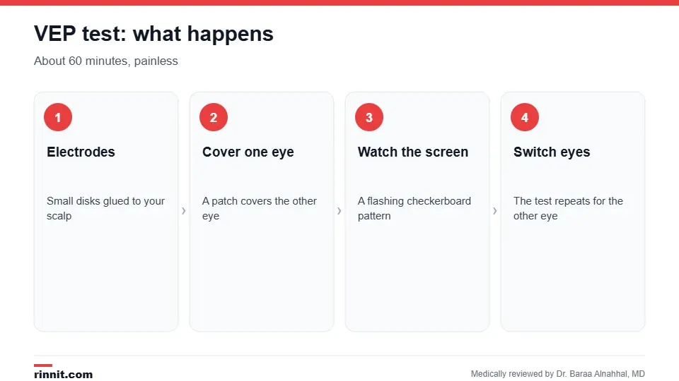
A VEP test takes up to 60 minutes.
Why is it done?

VEP checks the entire visual pathway.
Multiple sclerosis, the most common reason, because MS often damages the optic nerve.
Optic neuritis, inflammation of the optic nerve.
Optic nerve glioma, usually linked to neurofibromatosis type 1.
Pressure on the optic pathways, such as from a pituitary tumor or hydrocephalus.
Damage from head or brain injury.
Toxic optic neuropathy from certain medicines or toxins such as carbon monoxide.
Ischemic optic neuropathy and optic nerve damage from infections.
Monitoring known conditions such as MS, Leber optic neuropathy and Friedreich's ataxia. Sources: [1]
How do I prepare?
Wash your hair the night before or the morning of the test.
Don't use hair sprays, oils or gels, which can stop the electrodes from sticking.
Bring your glasses if you wear them, since you need to see the screen clearly.
Follow any other instructions your provider gives. Sources: [1]
What happens during the test?
A technician attaches small metal electrodes to specific spots on your scalp with removable paste.
A patch covers one eye.
You sit in front of a monitor and focus on a dot in the center of a flashing checkerboard pattern.
The computer records your brain's response over many repetitions.
The process is repeated for the other eye. Sources: [1]
The test takes up to 60 minutes. It isn't painful, and you can return to normal activities right away. Sources: [1]
Understanding your results
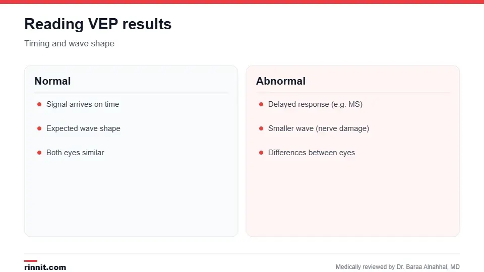
Timing and wave size tell different stories.
A specialist reads the wave patterns and sends a report to your provider. The most important measure is the P100 wave, the main peak about a tenth of a second after the pattern changes. A delayed signal suggests damage to the myelin coating of the optic nerve, typical of MS and optic neuritis. A smaller signal suggests loss of nerve fibers. Differences between the two eyes point to a problem in front of the optic chiasm. Your provider combines the results with your exam, a brain MRI and eye tests such as optical coherence tomography (OCT) and a visual field test. Sources: [1, 2, 3]
Risks
A VEP test is safe and noninvasive. There's no radiation and no needles. Some people find the flashing pattern tiring. Sources: [1]
Conclusion
A VEP test is a simple, painless way to check how well your visual pathway carries signals to your brain. It can detect optic nerve damage even when you don't notice vision changes, which makes it especially useful when MS is suspected.
Keep learning: read about multiple sclerosis and optic neuritis.
Frequently asked questions
What does a VEP test measure?
How quickly and strongly visual signals travel from your eyes to your brain.
Why is a VEP test done for MS?
MS often damages the optic nerve, which shows up as a delayed VEP signal.
Is a VEP test painful?
No. It's noninvasive and uses only scalp electrodes.
How long does a VEP test take?
Up to 60 minutes.
How should I prepare?
Wash your hair and avoid sprays, oils and gels.
Can I wear glasses during the test?
Yes. You should see the screen as clearly as possible.
What is the P100 wave?
The main peak of the VEP response, used to judge timing.
What does a delayed VEP mean?
It suggests damage to the myelin coating of the optic nerve.
Is VEP the same as an EEG?
It uses similar electrodes but records the brain's response to visual patterns.
Are there any risks?
No significant risks. There's no radiation or needles.
Related Reading
Evoked Potential Test: VEP, BAER and SSEP for MS and Nerve Problems explains the nerve-signal tests that include VEP, BAER and SSEP.
Optic Neuritis: Symptoms, Causes, and Treatment optic nerve inflammation that VEP can detect.
What Is Multiple Sclerosis? A Plain-Language Guide to Symptoms, Types, and How It's Treated the most common reason for a VEP test.
Electroencephalogram (EEG): Types, How to Prepare and What Results Mean a related test that records brain waves.
Ischemic Optic Neuropathy: Understanding Sudden Vision Loss and Optic Nerve Damage optic nerve damage from poor blood flow.
Understanding the Optic Nerve: Your Brain’s Visual Gateway the nerve that carries vision to the brain.
References
Cleveland Clinic. Visual Evoked Potential (VEP Test): Procedure & Results. Cleveland Clinic. Last updated January 25, 2023. https://my.clevelandclinic.org/health/diagnostics/24650-visual-evoked-potential-vep-test
Vidal-Jordana A, Rovira A, Arrambide G, et al. Optic nerve topography in multiple sclerosis diagnosis: the utility of visual evoked potentials. Neurology. 2021;96(4):e482-e490. PMID 33328323. https://doi.org/10.1212/WNL.0000000000011339
Filgueiras TG, Oyamada MK, Hokazono K, et al. Comparison of visual evoked potentials in patients affected by optic neuritis from multiple sclerosis or neuromyelitis optica spectrum disorder. J Neuroophthalmol. 2022;42(1):e32-e39. PMID 34348361. https://doi.org/10.1097/WNO.0000000000001285
Medical disclaimer
This article is for general education only and does not replace advice from a qualified healthcare professional. Talk to your doctor about your own diagnosis and treatment, and seek emergency care right away for severe or sudden symptoms.



Comments