Thoracoscopy: Types, How It's Done, Risks and Recovery

Updated: 2 days ago
Written and medically reviewed by Dr. Baraa Alnahhal, MD · Last reviewed: November 2026 · How we review
Quick Answer
Thoracoscopy is a minimally invasive procedure that uses a thin camera with a light, called a thoracoscope, to look inside the chest at the lungs and the space around them. Through small incisions between the ribs, providers can take biopsies, drain fluid and treat problems. It's usually done under general anesthesia, and full recovery typically takes 4–6 weeks.
What the research says
It has one of the highest diagnostic yields for fluid around the lungs: a network meta-analysis of 64 studies and 8,744 patients with unexplained exudative pleural effusions found rigid medical thoracoscopy had the highest diagnostic yield (95.0%), compared with 92.3% for semirigid thoracoscopy and 75.1% for closed (blind) pleural biopsy (Lyu S et al., European Respiratory Review, 2026) [2]
It's especially useful for cancer: a meta-analysis of 33 studies found medical thoracoscopy had 93% sensitivity and 100% specificity for diagnosing exudative pleural effusions, compared with 77% sensitivity for closed pleural biopsy, and performed better for malignancy (92% vs 72%) and mesothelioma (42% vs 26%) (Wei Y et al., Translational Lung Cancer Research, 2020) [3]
Key takeaways
A camera procedure to see inside the chest, take biopsies, drain fluid and treat lung or pleural problems.
Medical thoracoscopy (pleuroscopy) is done by a pulmonologist; VATS is surgery by a thoracic surgeon.
Usually two or three incisions under half an inch, with general anesthesia.
A chest tube typically stays in for one to two days after biopsies or drainage.
Normal activities resume within about two weeks; full recovery takes four to six weeks.
What is thoracoscopy?
The lungs are covered by a thin lining, the pleura, with a small space between the lung and chest wall. Imaging such as a chest X-ray, CT or ultrasound can show fluid or abnormal areas there, but not always their cause. Thoracoscopy lets providers look directly at the pleura and lung surface, take tissue samples and treat the problem through small incisions. Sources: [1]
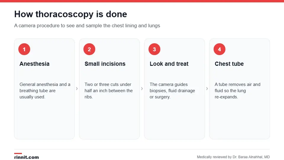
Thoracoscopy uses small incisions between the ribs.
Why is it done?
To find the cause of fluid around the lungs (pleural effusion) or pleurisy when other tests haven't.
To take biopsies of the pleura, lung or lymph nodes, such as for lung cancer or mesothelioma.
To drain fluid and prevent it from returning by sealing the pleural space (pleurodesis).
To treat a collapsed lung (pneumothorax) by removing air pockets.
To remove part of the lung, such as for cancer or lung volume reduction.
In cancers of the thymus or esophagus. Sources: [1]
Medical thoracoscopy vs VATS
Type | Who does it | Typical use |
Medical thoracoscopy (pleuroscopy) | Pulmonologist | Examining the pleura, biopsies, draining fluid, pleurodesis |
Thoracic surgeon | Lung resection, removing damaged tissue, more complex surgery |
Sources: [1]
How do I prepare?
Stop blood thinners a few days before, as your provider directs.
You may be asked not to eat or drink on the day of the procedure.
Arrange for someone to drive you home if you're discharged the same day. Sources: [1]
What happens during the procedure?
You receive general anesthesia through an IV, and a breathing tube is placed.
The surgeon makes two or three small incisions, each under half an inch, between the ribs.
The thoracoscope and instruments are inserted, and images appear on a monitor.
The provider examines the chest, takes samples or performs the planned treatment.
A chest tube is placed to remove air and fluid so the lung re-expands.
The incisions are closed and you're woken up. Sources: [1]
The procedure takes from about half an hour to several hours, depending on what's done. Sources: [1]
Thoracoscopy vs thoracotomy
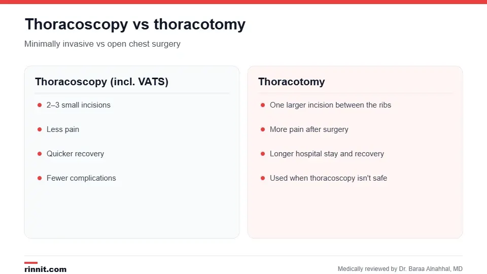
Thoracoscopy is less invasive than open chest surgery.
A thoracotomy opens the chest through a larger incision. Thoracoscopy is less invasive, with less pain, a quicker recovery and fewer complications, but a thoracotomy may still be needed if the procedure can't be done safely through small incisions. Sources: [1]
Recovery
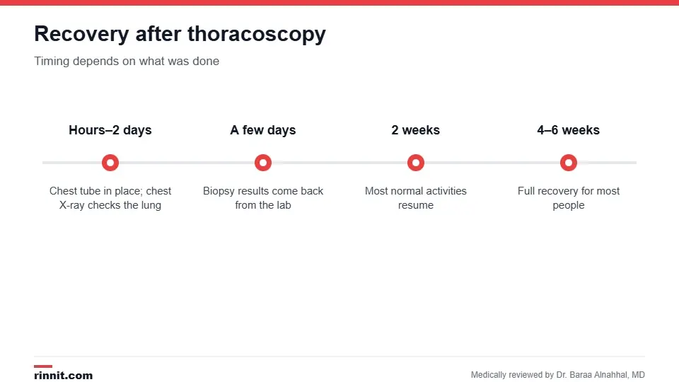
Recovery depends on what was done.
You may have a sore throat or hoarseness from the breathing tube, and pain at the incisions, which is treated with medicine.
A chest tube usually stays in for one to two days after biopsies or drainage, and a chest X-ray checks the lung.
Most people stay in the hospital while drains are in; some go home after a few hours.
Most normal activities resume within two weeks; full recovery usually takes four to six weeks. Sources: [1]
Risks
Complications occur in about 2–11% of procedures and can include bleeding, infection, fever, air leaks, a collapsed lung and fluid around the lung. The estimated risk of death is about 0.3% for medical thoracoscopy and slightly higher for VATS. Sources: [1]
Results
Your provider can tell you right away what they saw and whether the problem was treated. Biopsy results take a few days and show whether a spot is cancerous or benign; cancer results may include staging. Sources: [1, 2, 3]
After you go home, call your provider for trouble breathing, fever, chest pain or new swelling in your chest or neck. Sources: [1]
Conclusion
Thoracoscopy lets providers see and sample the inside of your chest without a large incision. It's one of the most accurate ways to find the cause of fluid around the lungs and can often treat the problem in the same procedure. Ask whether you'll have medical thoracoscopy or VATS, and what recovery to expect.
Frequently asked questions
What is thoracoscopy used for?
To look inside the chest, take biopsies, drain fluid and treat lung or pleural problems.
Is thoracoscopy surgery?
It can be diagnostic (medical thoracoscopy) or surgical (VATS).
Will I be asleep?
Usually yes; general anesthesia is typically used.
How long does it take?
From about half an hour to several hours, depending on what's done.
How big are the incisions?
Usually two or three, each under half an inch.
How long will the chest tube stay in?
Typically one to two days after biopsies or fluid drainage.
How long is recovery?
Most activities resume within two weeks; full recovery takes four to six weeks.
What are the risks?
Bleeding, infection, air leaks, collapsed lung and fluid build-up; complications occur in about 2–11%.
How accurate is thoracoscopy for pleural effusions?
Very accurate; studies show diagnostic yields of about 92–95%.
When will I get biopsy results?
Usually within a few days.
Related Reading
Video-Assisted Thoracic Surgery (VATS): Procedure and Benefits the surgical form of thoracoscopy.
Pleurisy: Complete Guide to Symptoms, Causes, and Treatment inflammation of the pleura that thoracoscopy can investigate.
What Is Mesothelioma? The Rare Cancer Linked to Asbestos — and What Treatment Looks Like a pleural cancer diagnosed by thoracoscopic biopsy.
Pneumothorax: Understanding Collapsed Lung Causes and Care a collapsed lung that may be treated with thoracoscopy.
Lung Cancer: Symptoms, Causes, Diagnosis, and Treatment Explained a cancer that thoracoscopy helps diagnose and treat.
References
Cleveland Clinic. Thoracoscopy: Procedure Details and Recovery. Cleveland Clinic. Last updated July 6, 2022. https://my.clevelandclinic.org/health/diagnostics/23463-thoracoscopy
Lyu S, Zheng Z, Liao L, et al. Diagnostic performance and safety of image-guided pleural biopsy and medical thoracoscopy for undiagnosed exudative pleural effusion: a systematic review and network meta-analysis. European Respiratory Review. 2026;35(180). PMID 42270119. https://doi.org/10.1183/16000617.0310-2025
Wei Y, Shen K, Lv T, et al. Comparison between closed pleural biopsy and medical thoracoscopy for the diagnosis of undiagnosed exudative pleural effusions: a systematic review and meta-analysis. Translational Lung Cancer Research. 2020;9(3):446-458. PMID 32676309. https://doi.org/10.21037/tlcr.2020.03.28
Medical disclaimer
This article is for general education only and does not replace advice from a qualified healthcare professional. Talk to your doctor about your own diagnosis and treatment, and seek emergency care right away for severe or sudden symptoms.



Comments