Video-Assisted Thoracic Surgery (VATS): Procedure and Benefits

Updated: 3 days ago
Medically reviewed by Dr. Baraa Alnahhal, MD · Last reviewed: October 2026
TL;DR: Video-assisted thoracic surgery (VATS) is a minimally invasive surgical technique used to diagnose and treat conditions within the chest cavity. [1] By using a small camera called a thoracoscope and specialized instruments through tiny incisions, surgeons can perform complex operations with less pain and faster recovery times than traditional open-chest surgery. [1] VATS is most commonly employed for lung cancer treatment, esophageal surgery, and managing various lung diseases.
Quick Answer: What is video-assisted thoracic surgery (VATS)?
Video-assisted thoracic surgery (VATS) is a minimally invasive procedure in which surgeons make small incisions between the ribs and insert a thoracoscope, a thin tube with a tiny camera, to view, diagnose and treat problems inside the chest. [1] It is often used for lung resections and biopsies, and it offers shorter hospital stays, less pain and faster recovery. [1]
What the research says
VATS lobectomy gave better early recovery than open surgery in a randomized trial. In the VIOLET trial (503 patients with known or suspected lung cancer), self-reported physical function at 5 weeks was better after VATS (mean difference 4.65, 95% CI 1.69–7.61), median hospital stay was 4 vs 5 days, and fewer VATS patients had complications before discharge (RR 0.74, 95% CI 0.66–0.84), with similar overall and progression-free survival (Lim et al., Health Technol Assess, 2022) [2].
Long-term survival did not differ significantly between VATS and open surgery. A 12.8-year median follow-up of a randomized trial that enrolled 206 patients with proven or suspected early-stage non-small-cell lung cancer found no significant differences in overall survival (P = 0.29), disease-free survival (P = 0.17), or cancer-specific survival (P = 0.31); the authors note that larger trials are needed to fully explore possible differences (Dittberner et al., Eur J Cardiothorac Surg, 2025) [3].
There is a trade-off: open surgery retrieved more lymph nodes than thoracoscopic surgery. A meta-analysis of 42 studies (8,947 participants) found a higher total lymph node count with thoracotomy than with minimally invasive thoracoscopic lobectomy (SMD −0.22, 95% CI −0.28 to −0.16), but no significant difference versus the robotic-assisted subgroup (SMD 0.12, 95% CI −0.04 to 0.27) (Liu et al., Medicine (Baltimore), 2025) [4].
Overview of VATS Technology
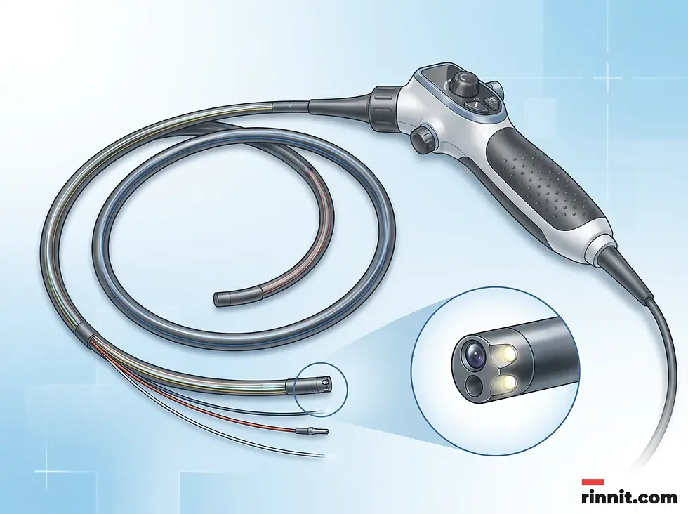
Video-assisted thoracic surgery represents a significant advancement in cardiothoracic medicine, allowing for complex internal procedures without the need for large, rib-spreading incisions. The core of this technology is the thoracoscope, which transmits high-definition images of the chest cavity to a video monitor in the operating room. [1] This visual guidance enables surgeons to perform precise movements using specialized instruments inserted through separate, quarter-inch to half-inch incisions. [1]
Surgeons may also utilize robotic-assisted VATS, where they control robotic arms through the same small incisions. [1] Whether traditional or robotic, the goal remains the same: to achieve the necessary surgical outcome while minimizing trauma to the patient's body. VATS is now used more and more often for many thoracic operations [1] because of its advantages for patients.

Conditions Treated and Surgical Types
While VATS is most widely recognized for its role in diagnosing and treating lung cancer, its applications extend to a wide variety of thoracic conditions. [1] Surgeons use VATS for both diagnostic purposes, such as performing biopsies, and therapeutic interventions, such as removing diseased tissue or organs. The following table outlines the primary types of VATS procedures and the conditions they address.
Procedure Type | Primary Use and Description |
Lung Resection | Removal of diseased lung tissue, ranging from small segments (wedge resection) to an entire lung (pneumonectomy). [1] |
Esophagectomy | Removal of all or part of the esophagus, typically to treat esophageal cancer. |
Thymectomy | Removal of the thymus gland, often performed for patients with myasthenia gravis or thymus tumors. |
Decortication | A procedure to remove infected material or restrictive scar tissue from the surface of the lung. [1] |
LVRS | Lung Volume Reduction Surgery, which removes damaged lung tissue to help the remaining lung function better in COPD patients. |
The VATS Procedure: Preparation and Execution
Preparation for VATS involves a comprehensive medical evaluation, including blood work, imaging tests like CT or PET scans, and pulmonary function testing to ensure the patient can tolerate the procedure. [1] Patients are typically required to fast for several hours beforehand and may be asked to adjust certain medications. [1] Surgeons may also ask patients to quit smoking. [1]
During the surgery, the patient is placed under general anesthesia and positioned on their nonsurgical side. [1] The surgeon makes one or more small incisions to insert the thoracoscope and instruments. A temporary chest tube is often placed at the end of the procedure to drain any excess air or fluid from the chest cavity. [1] Most lung cancer operations using VATS take between two and three hours [1], though duration varies based on complexity.
Benefits and Potential Risks
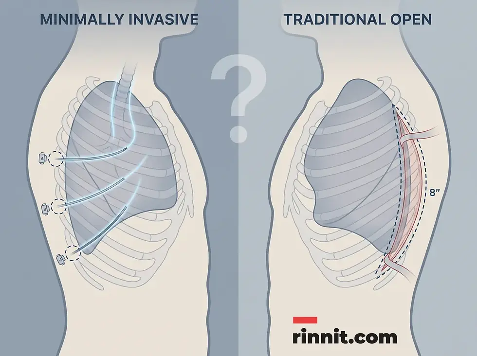
The primary advantage of VATS over traditional open-chest surgery (thoracotomy) is the reduction in physical trauma. Because the ribs are not spread apart, patients experience significantly less postoperative pain [1] [2] and have a lower risk of wound infections. However, like any major surgery involving vital organs, VATS does carry potential risks that patients should discuss with their surgical team.
Benefits of the VATS approach include shorter hospital stays, minimal scarring, and a faster return to normal respiratory function. [1] Potential complications, while less frequent than in open surgery, can include blood clots, pneumonia, collapsed lung (pneumothorax), or damage to nearby nerves and blood vessels. [1] Surgeons carefully weigh these factors when determining the best surgical approach for each individual patient.
Frequently Asked Questions
What is the main difference between VATS and a thoracotomy?
VATS is a minimally invasive procedure using small incisions and a camera, whereas a thoracotomy is a traditional open surgery requiring a large incision and spreading of the ribs to access the chest cavity.
How long is the recovery time after a VATS procedure?
Recovery time varies depending on the procedure, from about two weeks for some to eight weeks for others. Most people spend one or two nights in the hospital after surgery.
Is VATS used for all lung cancer surgeries?
VATS is a common choice for lung cancer resections, but the decision depends on the size and location of the tumor. Some complex cases may still require a traditional open approach.
What is a thoracoscope?
A thoracoscope is a thin tube equipped with a tiny video camera and light source. It allows the surgeon to see high-definition images of the inside of the chest on a monitor.
Will I have a chest tube after VATS?
Yes, most patients have a temporary chest tube placed at the end of the surgery to drain air or fluid from the chest cavity. It is usually removed before you go home.
What kind of anesthesia is used for VATS?
VATS is performed under general anesthesia, meaning you will be completely asleep and will not feel any pain during the procedure.
Are there scars after VATS?
Because the incisions are very small (typically quarter-inch to half-inch), the resulting scars are minimal and much less noticeable than the large scar from a thoracotomy.
Can VATS be used for diagnosis?
Yes, VATS is frequently used for diagnostic purposes, such as taking a biopsy of a lung nodule or lymph node to check for cancer or infection.
What are the risks of VATS?
Potential risks include infection, bleeding, blood clots, pneumonia, and a collapsed lung (pneumothorax). However, these complications occur less frequently than with open surgery.
How do I prepare for VATS?
Preparation involves fasting, blood tests, imaging scans, and potentially stopping certain medications or quitting smoking as directed by your surgeon.
Related Reading
Learn more in our guide to Bullectomy.
Thoracoscopy: Types, How It's Done, Risks and Recovery explains the camera procedure used to see inside the chest and sample the pleura.
References
Cleveland Clinic. Video-Assisted Thoracoscopic Surgery (VATS). Cleveland Clinic. Last updated 04/17/2025. https://my.clevelandclinic.org/health/treatments/17617-video-assisted-thoracic-surgery-vats
Lim E, Harris RA, McKeon HE, et al. Impact of video-assisted thoracoscopic lobectomy versus open lobectomy for lung cancer on recovery assessed using self-reported physical function: VIOLET RCT. Health Technol Assess. 2022;26(48):1-162. PMID 36524582. https://doi.org/10.3310/THBQ1793
Dittberner FA, Bendixen M, Licht PB. Long-term survival from a randomized controlled trial of lobectomy by video-assisted thoracoscopic surgery versus thoracotomy for early-stage lung cancer. Eur J Cardiothorac Surg. 2025;67(7):ezaf017. PMID 39873729. https://doi.org/10.1093/ejcts/ezaf017
Liu Y, Sun Z, Deng Y, Li H, Li Y, Lin D. Thoracotomy is better than minimally invasive thoracoscopic lobectomy in the lymph node dissection of lung cancer: a systematic review and meta-analysis. Medicine (Baltimore). 2025;104(46):e45657. PMID 41239607. https://doi.org/10.1097/MD.0000000000045657
Medical disclaimer
This article is for educational purposes only and is not a substitute for professional medical advice, diagnosis, or treatment. Always seek the advice of your physician or other qualified health provider with any questions about a medical condition.


Comments