MRI-Guided Prostate Biopsy: Fusion Biopsy, Prep, Risks and Results

Updated: 3 days ago
Written and medically reviewed by Dr. Baraa Alnahhal, MD · Last reviewed: November 2026 · How we review
Quick Answer
An MRI-guided prostate biopsy uses magnetic resonance imaging (MRI) to find suspicious areas in the prostate and guide needles to sample exactly those areas for signs of prostate cancer. It's used for a high PSA, an abnormal rectal exam or a suspicious MRI. Compared with a standard biopsy, it finds more clinically significant cancers and fewer harmless ones.
What the research says
MRI targeting finds more significant cancers: in 500 men, MRI-targeted biopsy detected clinically significant prostate cancer in 38% vs. 26% with standard biopsy, and 28% of men in the MRI group avoided biopsy altogether (Kasivisvanathan V et al., New England Journal of Medicine, 2018) [2]
MRI is more sensitive than standard biopsy: in 576 men who had both, multiparametric MRI detected 93% of clinically significant cancers vs. 48% for standard ultrasound biopsy, and MRI triage could let about 27% of men avoid a first biopsy (Ahmed HU et al., Lancet, 2017) [3]
Key takeaways
MRI pinpoints suspicious areas so needles can target them.
Fusion biopsy combines MRI images with live ultrasound.
It's done as an outpatient with local anesthetic.
Samples are taken through the rectum or the perineum.
Mild bleeding in urine or semen for a few days is common.
What is an MRI-guided prostate biopsy?
A standard prostate biopsy takes samples from set areas of the prostate using ultrasound, which can miss some cancers. A multiparametric MRI (mpMRI) first shows areas that look suspicious. During the biopsy, software overlays the MRI on the live transrectal ultrasound image, so the urologist can aim needles precisely. A pathologist then examines the samples. MRI alone can't rule out cancer, so a biopsy is still needed to confirm it. Sources: [1]
Why it's done

Targeting helps find cancers that matter.
Elevated PSA, an abnormal digital rectal exam or a suspicious MRI.
PSA that keeps rising after a negative standard biopsy.
Monitoring known low-risk cancer during active surveillance.
Planning focal therapy.
Ruling out benign conditions such as an enlarged prostate or prostatitis. Sources: [1]
Transrectal vs. transperineal approach
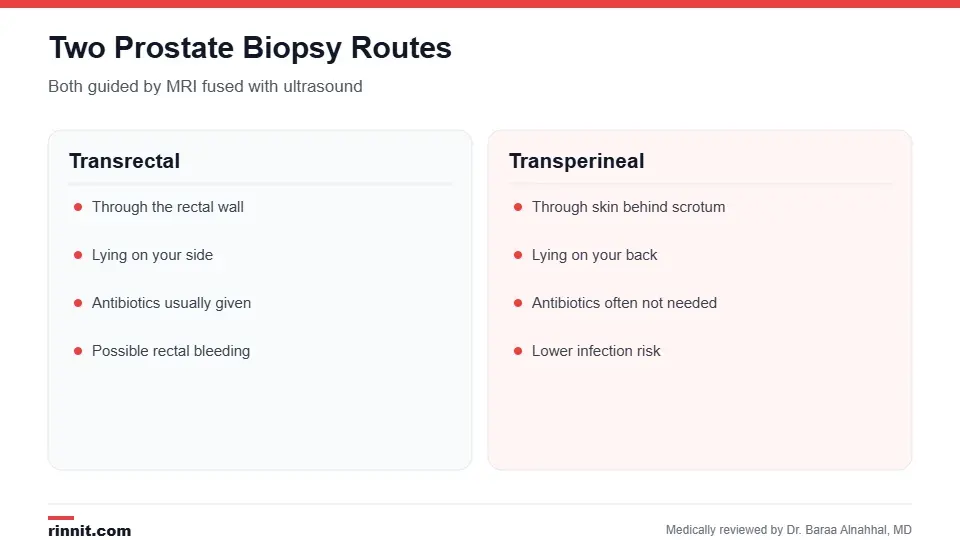
Your urologist chooses the approach.
Approach | How samples are taken | Position |
Transrectal | Needle passes through the rectal wall | Lying on your side |
Transperineal | Needle passes through the skin between the anus and scrotum | Lying on your back with legs in stirrups |
Antibiotics are usually given for transrectal biopsies and are often unnecessary for transperineal ones. Sources: [1]
How to prepare
Tell your provider about implanted metal devices, such as a pacemaker or joint replacement, before the MRI.
Empty your bowels; an enema may be suggested.
Stop NSAIDs and blood thinners as instructed.
Take antibiotics before and after if prescribed.
Arrange a ride if you'll have a sedative. Sources: [1]
What happens during the procedure

You can usually go home the same day.
You have an mpMRI, sometimes with IV contrast, before the biopsy.
On biopsy day, the area is numbed with local anesthetic; a mild sedative may be given.
The ultrasound probe is placed, and the MRI images are fused with live ultrasound.
Needles take samples from the targeted areas and standard areas, typically 12 to 16 cores.
The samples go to a pathologist. Sources: [1]
You'll feel pressure, but it shouldn't be painful. Sources: [1]
Risks and recovery
You can go home once any sedation wears off. Expect mild discomfort and some blood in your urine or semen for a few days, plus rectal bleeding after a transrectal biopsy. Possible risks include infection, bleeding, bruising and, rarely, an allergic reaction to MRI contrast. Call your provider for fever or chills. Sources: [1]
Results
Results usually take about a week. They may be negative, positive or suspicious. If cancer is found, the report includes the Gleason score, which helps guide treatment. False negatives are possible, so your provider may recommend continued PSA monitoring. Sources: [1]
Conclusion
An MRI-guided prostate biopsy aims needles at the areas most likely to hold cancer. This targeted approach finds more cancers that need treatment, helps avoid overdiagnosis of harmless ones and may spare some men an unnecessary biopsy.
Keep learning: read about the PSA test and prostate ultrasound and biopsy.
Frequently asked questions
What is an MRI-guided prostate biopsy?
A biopsy that uses MRI to target suspicious areas of the prostate.
What is a fusion biopsy?
A biopsy that combines MRI images with live ultrasound.
Why is it better than a standard biopsy?
It finds more significant cancers and fewer harmless ones.
Is it painful?
You'll feel pressure, but local anesthetic prevents pain.
How many samples are taken?
Usually 12 to 16.
Do I need antibiotics?
Usually for transrectal biopsies; often not for transperineal ones.
Is blood in my urine normal afterward?
Yes, for a few days.
When will I get results?
Usually in about a week.
Can MRI alone diagnose prostate cancer?
No. A biopsy is needed to confirm it.
What should I watch for afterward?
Fever, chills or heavy bleeding.
Related Reading
PSA Test: Normal Levels by Age, How to Prepare and What High Results Mean the blood test that often leads to a biopsy.
Prostate Ultrasound and Biopsy: Why It's Done, How to Prepare, What to Expect and Recovery the standard ultrasound-guided biopsy.
Gleason Score: How Prostate Cancer Is Graded and What Your Score Means how prostate cancer is graded.
PSMA PET Scan for Prostate Cancer: How It Works, How to Prepare and How Accurate It Is an imaging test for prostate cancer spread.
Focal Therapy for Prostate Cancer a targeted treatment option.
References
Cleveland Clinic. MRI-Guided Prostate Biopsy: How It Works. Cleveland Clinic. Last updated April 9, 2023. https://my.clevelandclinic.org/health/diagnostics/16382-mri-guided-prostate-biopsy
Kasivisvanathan V, Rannikko AS, Borghi M, et al. MRI-targeted or standard biopsy for prostate-cancer diagnosis. New England Journal of Medicine. 2018;378(19):1767-1777. PMID 29552975. https://doi.org/10.1056/NEJMoa1801993
Ahmed HU, El-Shater Bosaily A, Brown LC, et al. Diagnostic accuracy of multi-parametric MRI and TRUS biopsy in prostate cancer (PROMIS): a paired validating confirmatory study. Lancet. 2017;389(10071):815-822. PMID 28110982. https://doi.org/10.1016/S0140-6736(16)32401-1
Medical disclaimer
This article is for general education only and does not replace advice from a qualified healthcare professional. Talk to your doctor about your own diagnosis and treatment, and seek emergency care right away for severe or sudden symptoms.



Comments