Magnetoencephalography (MEG): Mapping the Brain Before Epilepsy Surgery

Updated: 2 days ago
Written and medically reviewed by Dr. Baraa Alnahhal, MD · Last reviewed: November 2026 · How we review
Quick Answer
Magnetoencephalography (MEG) is a painless, noninvasive test that records the tiny magnetic fields produced by the brain's electrical activity. It's mainly used to find where seizures start before epilepsy surgery and to map movement, sensation, language and vision areas before brain tumor surgery. It uses no radiation and no needles. The test takes about 1 to 2 hours.
What the research says
MEG improves epilepsy surgery planning: in 1,000 people evaluated for epilepsy surgery over 28 years, removing the brain area MEG identified was linked to higher chances of long-term seizure freedom, with especially strong accuracy in epilepsy starting outside the temporal lobe (Rampp S et al., Brain, 2019) [2]
MEG findings predict surgical success: in 132 people who had epilepsy surgery, 85% of those whose MEG findings matched the area removed became seizure-free, compared with only 37% of those with nonspecific or mismatched MEG findings (Englot DJ et al., Epilepsia, 2015) [3]
Key takeaways
Records the brain's magnetic fields with a sensor helmet.
Finds where seizures start and maps key brain areas.
Painless, no radiation and no needles.
Remove all metal; tell your provider about implants.
Takes about 1–2 hours; results may take days to weeks.
How does MEG work?
Nerve cells create tiny electrical currents, and these currents produce weak magnetic fields. MEG uses extremely sensitive sensors called SQUIDs (superconducting quantum interference devices) inside a helmet to detect them. The signals are combined with a brain MRI to make a functional map, called magnetic source imaging. Sources: [1]
Why is it done?

MEG pinpoints brain activity in space and time.
To locate where seizures begin in people with epilepsy, especially focal seizures, to plan epilepsy surgery.
To map movement, sensation, language and vision areas before brain tumor surgery.
For research on brain function. Sources: [1, 2, 3]
MEG is often combined with other tests, such as SEEG, when the seizure source is unclear. Sources: [1]
MEG compared with other tests
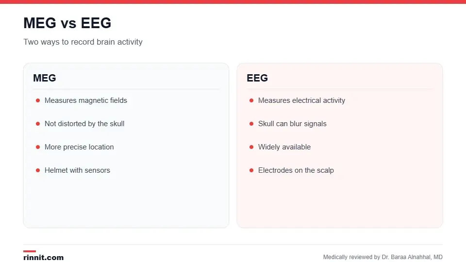
MEG signals aren't distorted by the skull.
Test | What it measures | Strength |
MEG | Magnetic fields from brain activity | Precise location and timing |
Electrical activity on the scalp | Widely available, but the skull can blur signals | |
Blood flow changes during tasks | Shows general active areas | |
MRI | Brain structure | Detailed anatomy |
Sources: [1]
How do I prepare?
Follow instructions about eating, drinking and medicines.
Don't wear makeup or hair products.
Remove all metal, including jewelry, glasses, hairpins and underwire bras.
Tell your provider about implants, such as pacemakers, defibrillators, cochlear implants, nerve stimulators, metal plates or programmable shunts, which may interfere with the test. Sources: [1]
What happens during the test?
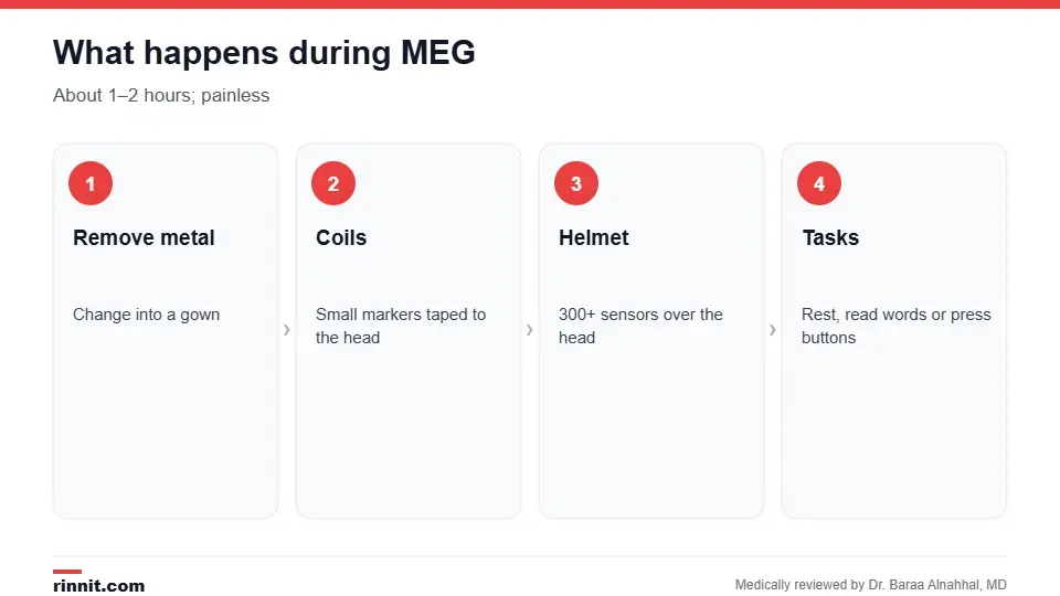
MEG takes about one to two hours.
You change into a gown and remove all metal.
Small positioning coils are taped to your head and their locations measured.
You lie on a table or sit in a chair, and the sensor helmet is placed over your head.
You keep your head still and may rest, read words, answer questions or press a button; a small, harmless electrical pulse may be applied to a finger.
Staff can see and talk with you through video and an intercom. Sources: [1]
The test takes about one to two hours, and you may even fall asleep. Young children may need sedation. Sources: [1]
Risks
MEG has no known risks. It doesn't use radiation. If sedation was used, you'll be watched for a short time and need someone to drive you home. Sources: [1]
Understanding your results
Specialists may take days or weeks to review the recordings, because MEG often guides complex surgical decisions. Your care team will explain what the results mean and the next steps. Sources: [1]
Conclusion
MEG gives a precise, noninvasive look at brain activity. It's especially valuable for people considering epilepsy or brain tumor surgery, helping surgeons target the right area and protect important functions.
Keep learning: read about epilepsy surgery and the EEG.
Frequently asked questions
What is MEG?
A test that records the brain's magnetic fields to map activity.
Why is it done?
Mainly to find where seizures start and map brain areas before surgery.
Does it hurt?
No. It's painless and noninvasive.
Is there radiation?
No.
How long does it take?
About one to two hours.
Can I have MEG with a pacemaker?
Tell your provider; metal implants may prevent testing.
Can I fall asleep during MEG?
Yes, sometimes that's fine.
How is MEG different from EEG?
MEG measures magnetic fields and isn't distorted by the skull.
When will I get results?
It may take days or weeks.
Do children need sedation?
Infants and young children sometimes do.
Related Reading
Epilepsy Surgery: Types, Tests, Risks, Recovery, and Success Rates surgery planned with MEG.
Electroencephalogram (EEG): Types, How to Prepare and What Results Mean the electrical brain test often done alongside.
SEEG (Stereoelectroencephalography): Finding Where Seizures Start a deep-brain electrode test for seizures.
Functional MRI (fMRI): What It Shows, How It Works and How to Prepare another way to map brain function.
Epilepsy: Seizures, Symptoms, Causes, and Treatment the main condition MEG evaluates.
References
Cleveland Clinic. Magnetoencephalography (MEG): What It Is, Purpose & Uses. Cleveland Clinic. Last updated March 30, 2023. https://my.clevelandclinic.org/health/diagnostics/17218-magnetoencephalography-meg
Rampp S, Stefan H, Wu X, et al. Magnetoencephalography for epileptic focus localization in a series of 1000 cases. Brain. 2019;142(10):3059-3071. PMID 31373622. https://doi.org/10.1093/brain/awz231
Englot DJ, Nagarajan SS, Imber BS, et al. Epileptogenic zone localization using magnetoencephalography predicts seizure freedom in epilepsy surgery. Epilepsia. 2015;56(6):949-958. PMID 25921215. https://doi.org/10.1111/epi.13002
Medical disclaimer
This article is for general education only and does not replace advice from a qualified healthcare professional. Talk to your doctor about your own diagnosis and treatment, and seek emergency care right away for severe or sudden symptoms.



Comments