Functional MRI (fMRI): What It Shows, How It Works and How to Prepare

Updated: 2 days ago
Written and medically reviewed by Dr. Baraa Alnahhal, MD · Last reviewed: November 2026 · How we review
Quick Answer: What is a functional MRI?
A functional MRI (fMRI) uses an MRI machine to track blood flow in the brain while you do simple tasks, such as tapping your fingers. Areas that are working harder use more oxygen and show up brighter. Its main use is to map important brain areas before surgery or radiation so they can be protected. [1]
What the research says
fMRI agrees well with the older invasive language test. Across 23 studies with 442 patients, fMRI identified atypical language dominance with a sensitivity of 83.5% and a specificity of 88.1% compared with the Wada test, an invasive test that puts one side of the brain to sleep. (Dym et al., Radiology, 2011) [2]
Results in children look promising. In a 2025 meta-analysis of children having language mapping, fMRI and the Wada test agreed in 81% of cases, though confirmation may still be needed when language is not clearly on the left side. (de Macedo et al., Neuroradiology, 2025) [3]
Key takeaways
fMRI uses the same machine as a standard MRI but shows brain activity, not just structure. [1]
You do simple tasks during the scan so your care team can see which areas light up. [1]
It is mainly used to plan surgery or radiation for epilepsy and brain tumors. [1]
It does not use radiation, and its risks are similar to a standard MRI. [1]
The scan usually takes up to one hour. [1]
What is a functional MRI (fMRI)?
A functional MRI tracks blood flow in your brain to show which areas are working hardest at a given moment. Brain cells that are active use more oxygen, so those areas appear brighter on the scan. [1]
A standard MRI uses a powerful magnet, radio waves and a computer to make detailed 3D pictures of the inside of your body, without radiation. An fMRI uses the same type of machine, but it adds tasks that you do during the scan. [1]
How is fMRI different from a standard MRI?

Both tests use the same scanner. The difference is what they show.
Feature | Standard MRI | Functional MRI |
What it shows | Brain structure | Which brain areas are active |
What you do | Lie still | Lie still and do simple tasks |
Main use | Diagnosing many conditions | Mapping the brain before surgery or radiation |
Radiation | None | None |
Sources: [1]
fMRI is not common in everyday healthcare, and smaller facilities may not offer it. [1]
Why would you need an fMRI?
fMRI is mainly used to plan brain surgery and similar procedures. It helps surgeons avoid areas that control critical functions. You may need one before: [1]
Surgery for epilepsy
Removal of a brain tumor
Precision radiation therapy to the brain
Other brain procedures
Providers may also use it after treatment to check for changes in brain function. Researchers are studying other uses, and there is some evidence it may help with biofeedback therapy. [1]
How do you prepare for an fMRI?

Your care team will give you instructions. In general: [1]
Avoid caffeine on the day of your scan.
Follow instructions about your usual medicines.
Remove all metal, including jewelry, piercings, hearing aids and electronics. The magnet is strong enough to make metal dangerous.
Tell your provider if you have: [1]
A pacemaker, stimulator, joint replacement, cochlear implant or other implanted device
Metal fragments in your body
A medication patch or an insulin pump
A pregnancy, or think you might be pregnant
Trouble lying on your back for the full scan
Claustrophobia (fear of tight spaces). Anti-anxiety medicine may be an option.
Children may need medicine to help them relax. [1]
What happens during an fMRI?
You lie on a table that slides into a ring-shaped scanner. Your head rests in a helmet-like head coil that helps produce clearer images. Sound-canceling headphones block the machine's noise and let staff talk with you. [1]
Tasks during the scan
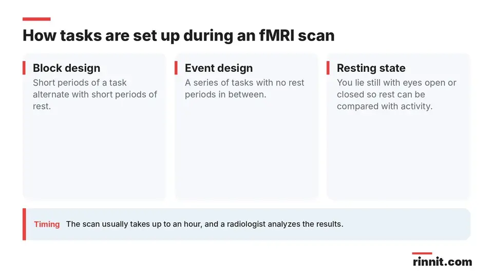
Tasks are arranged in one of two ways, and a resting scan may be added: [1]
Block design: short periods of a task alternate with short periods of rest.
Event design: a series of tasks with no rest periods in between.
Resting state: you lie still with your eyes open or closed so rest can be compared with activity.
Some scans use gadolinium contrast given through an IV. The scan generally takes up to an hour, and it can take longer if more data are needed. [1]
What happens after an fMRI?
You can usually go home right away. If you took anti-anxiety medicine, you will need someone to drive you. A radiologist analyzes the scan and sends the results to you and your referring provider, such as a neurologist or neurosurgeon. [1]
What are the benefits and risks?
fMRI is especially useful for planning surgery. It is usually preferred over other brain-mapping tests, which tend to be more invasive, use medicines with more side effects or map brain areas less accurately. [1]
Its main limitation is timing. The scan captures data second by second, but brain activity changes in tiny fractions of a second, so very fast changes cannot be detected. [1]
Risks are minimal and similar to a standard MRI. Side effects from gadolinium are rare. The main safety concern is metal in or on your body. [1]
fMRI vs. EEG
An EEG records brain activity as waves, while an fMRI shows similar activity as images. [1]
Conclusion
A functional MRI turns brain activity into a map. That map helps your surgical team plan the safest route around the areas you need for movement and other critical functions.
Preparation is mostly about metal safety and telling your team about implants, pregnancy or anxiety in tight spaces. Ask your provider what tasks you will be asked to do so you can practice beforehand.
Keep learning: Read our MRI guide or our guide to epilepsy surgery. For more background, see the RadiologyInfo page on functional MRI of the brain.
FAQ
What is a functional MRI?
It is an MRI scan that tracks blood flow in your brain to show which areas are most active while you do simple tasks. [1]
How is fMRI different from a regular MRI?
A standard MRI shows structure, while fMRI shows activity by having you do tasks during the scan. [1]
Why is fMRI done?
Mainly to map the brain before surgery or radiation for epilepsy or brain tumors so critical areas can be avoided. [1]
What tasks do you do during an fMRI?
Simple tasks such as tapping your fingers, done in blocks with rest or as a series without rest. [1]
How long does an fMRI take?
Generally up to an hour, sometimes longer if more data are needed. [1]
How do I prepare for an fMRI?
Avoid caffeine on the day of the scan and remove all metal, including jewelry and electronics. [1]
Does an fMRI use radiation?
No. It uses a magnet, radio waves and a computer, like a standard MRI. [1]
Is fMRI safe?
Risks are minimal and similar to a standard MRI; metal safety is the main concern. [1]
Can children have an fMRI?
Yes. Adults and children can have one, and children may need medicine to relax. [1]
What is the difference between fMRI and EEG?
An EEG records brain activity as waves, while fMRI shows similar activity as images. [1]
Related Reading
Epilepsy: Seizures, Symptoms, Causes, and Treatment explains the condition for which fMRI most often guides surgery.
Brain Cancer (Brain Tumors): Types, Symptoms, and Treatment Explained covers tumors that may need brain mapping before removal.
Brain AVM (Arteriovenous Malformation): Symptoms, Causes, and Treatment describes another brain condition where treatment is planned around critical areas.
Epilepsy Surgery: Types, Tests, Risks, Recovery, and Success Rates shows where fMRI fits in the surgical workup.
Magnetoencephalography (MEG): Mapping the Brain Before Epilepsy Surgery explains the magnetic brain scan used to find where seizures start.
Wada Test: Finding Language and Memory Before Brain Surgery explains the test that shows which side of the brain controls language and memory.
References
Cleveland Clinic. fMRI (Functional MRI): What It Is, Purpose, Procedure & Results. Cleveland Clinic. Last updated May 27, 2023. https://my.clevelandclinic.org/health/diagnostics/25034-functional-mri-fmri
Dym RJ, Burns J, Freeman K, et al. Is functional MR imaging assessment of hemispheric language dominance as good as the Wada test?: a meta-analysis. Radiology. 2011;261(2):446-455. PMID 21803921. https://doi.org/10.1148/radiol.11101344
de Macedo BA, Verdan S, Joshi DK, et al. Is functional MRI a reliable surrogate for the Wada test for preoperative assessment of hemispheric language laterality in pediatric patients? A systematic review and meta-analysis. Neuroradiology. 2025;67(12):3483-3494. PMID 41258566. https://doi.org/10.1007/s00234-025-03841-5
Medical disclaimer
This article is for general education only and does not replace advice from a qualified healthcare professional. Talk to your doctor about your own diagnosis and treatment, and seek emergency care right away for severe or sudden symptoms.



Comments