SEEG (Stereoelectroencephalography): Finding Where Seizures Start

Updated: 3 days ago
Written and medically reviewed by Dr. Baraa Alnahhal, MD · Last reviewed: November 2026 · How we review
Quick Answer
Stereoelectroencephalography (SEEG) is a minimally invasive procedure that places thin electrodes deep inside the brain through tiny holes in the skull to find exactly where drug-resistant seizures begin. You then stay in an Epilepsy Monitoring Unit, usually about a week, while seizures are recorded. Results show whether epilepsy surgery or another treatment could help. Serious complications are uncommon.
What the research says
SEEG has a low complication rate: a systematic review and meta-analysis of SEEG studies found an overall surgical complication rate of about 1.3%, with bleeding in about 1.0% and infection in about 0.8% of patients (Mullin JP et al., Epilepsia, 2016) [2]
SEEG compares well with older grid electrodes: in 260 procedures at one center, there were no clinically relevant complications after SEEG, compared with 7 bleeding events and 3 infections after subdural grids, and 76% of SEEG patients who went on to surgery or ablation had good seizure outcomes at one year (Tandon N et al., JAMA Neurol, 2019) [3]
Key takeaways
Finds where seizures start deep in the brain using implanted electrodes.
For drug-resistant focal epilepsy when other tests are unclear.
Placement takes about 4–6 hours under general anesthesia.
The hospital stay averages about one week while seizures are recorded.
Results guide epilepsy surgery, ablation or neurostimulation.
What is SEEG?
A standard EEG records brain activity from the scalp, which can miss seizures that start deep in the brain. SEEG, also called stereotactic EEG, places electrodes directly into deeper areas. Computers then build 3D maps showing where seizures start and how they spread. Sources: [1]
Who needs SEEG?

SEEG pinpoints where seizures begin.
Adults and children over 2 with drug-resistant (refractory) epilepsy, meaning at least two antiseizure medications haven't controlled seizures.
People with focal seizures when EEG and brain MRI results are unclear.
People being evaluated for epilepsy surgery.
To map movement, sensation and language areas before brain surgery.
To guide treatments such as laser interstitial thermal therapy (LITT) and responsive neurostimulation. Sources: [1]
SEEG isn't used for generalized epilepsy, and EEG is usually preferred for children under 2. Sources: [1]
How do I prepare?
You may need blood tests, an EKG and CT or MRI scans beforehand. Ask your care team about hair shaving and whether your seizure medicines will be lowered during monitoring. Sources: [1]
What happens during SEEG?
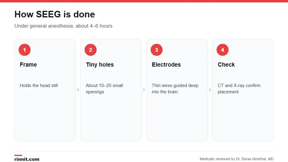
Electrodes are placed under general anesthesia.
You receive general anesthesia, so you feel no pain.
A rigid frame holds your head still.
About 10–20 tiny holes, roughly the width of a spaghetti noodle, are made in the skull.
Thin, flexible electrodes are guided to areas where seizures may start.
The frame is removed, your head is bandaged, and a CT scan and X-ray confirm electrode positions. Sources: [1]
In the Epilepsy Monitoring Unit
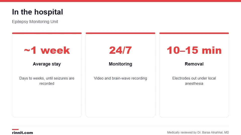
The stay lasts until enough seizures are recorded.
Staff watch you around the clock while equipment records your brain activity. The stay lasts from a few days to several weeks, depending on how often seizures happen, with an average of about one week. Removing the electrodes takes about 10–15 minutes under local anesthesia. Sources: [1]
Risks
SEEG is considered safe, but like any brain procedure it carries risks of bleeding in the brain, infection and stroke. Research suggests serious complications are uncommon. Sources: [1, 2, 3]
Recovery and results
Recovery usually takes about 24–48 hours. In most cases, SEEG finds the source of seizures. Your epilepsy team reviews the recordings to decide whether you can have surgery or other treatment; when surgery is recommended, it most often happens four to eight weeks later. Sources: [1]
Conclusion
SEEG gives a precise map of where seizures start, which can open the door to surgery or other treatments for people whose epilepsy hasn't responded to medicine. Its minimally invasive approach means fewer complications than older methods.
Keep learning: read about epilepsy surgery and the EEG.
Frequently asked questions
What is SEEG?
A procedure that places electrodes deep in the brain to find where seizures start.
Who needs SEEG?
People with drug-resistant focal epilepsy when other tests are unclear.
Is SEEG painful?
Placement is done under general anesthesia, so you feel no pain.
How long does placement take?
About 4–6 hours.
How long will I stay in the hospital?
Usually about a week, but it can range from days to weeks.
How many electrodes are placed?
Typically through about 10–20 tiny holes.
What are the risks?
Bleeding, infection and stroke, though serious problems are uncommon.
How is SEEG different from EEG?
EEG records from the scalp; SEEG records from deep inside the brain.
How are the electrodes removed?
In about 10–15 minutes with local anesthesia.
What happens after SEEG?
Your team decides on surgery or other treatment, often within four to eight weeks.
Related Reading
Epilepsy: Seizures, Symptoms, Causes, and Treatment the condition SEEG helps evaluate.
Epilepsy Surgery: Types, Tests, Risks, Recovery, and Success Rates the surgery SEEG often leads to.
Electroencephalogram (EEG): Types, How to Prepare and What Results Mean the scalp test usually done first.
Focal Seizure: Symptoms, Causes, and Management of Partial Seizures seizures that start in one area of the brain.
Responsive Neurostimulation (RNS) for Epilepsy: How It Works, Surgery and Results a device treatment guided by SEEG.
Magnetoencephalography (MEG): Mapping the Brain Before Epilepsy Surgery explains the magnetic brain scan used to find where seizures start.
References
Cleveland Clinic. Stereoelectroencephalography (SEEG): What It Is & Procedure. Cleveland Clinic. Last updated August 23, 2023. https://my.clevelandclinic.org/health/diagnostics/17457-seeg-test
Mullin JP, Shriver M, Alomar S, et al. Is SEEG safe? A systematic review and meta-analysis of stereo-electroencephalography-related complications. Epilepsia. 2016;57(3):386-401. PMID 26899389. https://doi.org/10.1111/epi.13298
Tandon N, Tong BA, Friedman ER, et al. Analysis of morbidity and outcomes associated with use of subdural grids vs stereoelectroencephalography in patients with intractable epilepsy. JAMA Neurol. 2019;76(6):672-681. PMID 30830149. https://doi.org/10.1001/jamaneurol.2019.0098
Medical disclaimer
This article is for general education only and does not replace advice from a qualified healthcare professional. Talk to your doctor about your own diagnosis and treatment, and seek emergency care right away for severe or sudden symptoms.



Comments