Liver Ultrasound: Why It's Done, How to Prepare, Types and What Results Mean

Updated: 2 days ago
Written and medically reviewed by Dr. Baraa Alnahhal, MD · Last reviewed: November 2026 · How we review
Quick Answer
A liver ultrasound is an imaging test that uses sound waves to create pictures of the liver, its blood vessels, the gallbladder and bile ducts. It's painless, uses no radiation and takes about 30 minutes. You usually fast for 6–12 hours beforehand. It can show fatty liver, scarring (cirrhosis), cysts, tumors and blocked ducts.
What the research says
Ultrasound measures liver fat almost as well as MRI: a meta-analysis of 38 studies and 5,056 people found ultrasound attenuation imaging detected any liver fat (steatosis grade S1 or higher) with an area under the curve of 0.97, close to MRI-PDFF at 0.99, while the controlled attenuation parameter scored 0.95; all methods were weaker for the most severe fat grade (Wu S et al., Academic Radiology, 2025) [2]
Ultrasound misses many early liver cancers: across 9 studies and 1,914 patients under surveillance, ultrasound detected early hepatocellular carcinoma with only 47% sensitivity (specificity 91%), whereas abbreviated MRI reached 83% sensitivity and 96% specificity, which is why some high-risk patients are offered MRI-based surveillance (Liu J et al., Clinical and Translational Oncology, 2026) [3]
Key takeaways
Fast for 6–12 hours so bowel gas is reduced and the gallbladder is full.
The sonographer scans the right upper abdomen while you hold your breath; Doppler adds blood-flow information.
Types include standard B-mode, Doppler, elastography (stiffness), attenuation imaging (fat) and contrast-enhanced ultrasound.
Results usually take a few days; a radiologist's report goes to your provider.
Limitations: bowel gas, body habitus and small early cancers can be missed, so MRI or CT is sometimes needed.
What is a liver ultrasound?
A transducer sends high-frequency sound waves into the abdomen and listens for the echoes. Different tissues reflect sound differently, so the computer can build a live, moving image. For the liver, the exam also covers the gallbladder, bile ducts, portal vein and hepatic veins, and often the spleen and right kidney. Sources: [1]
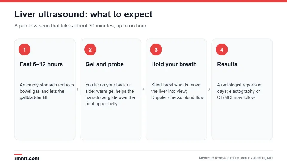
A liver ultrasound is painless and takes about 30 minutes.
Types of liver ultrasound
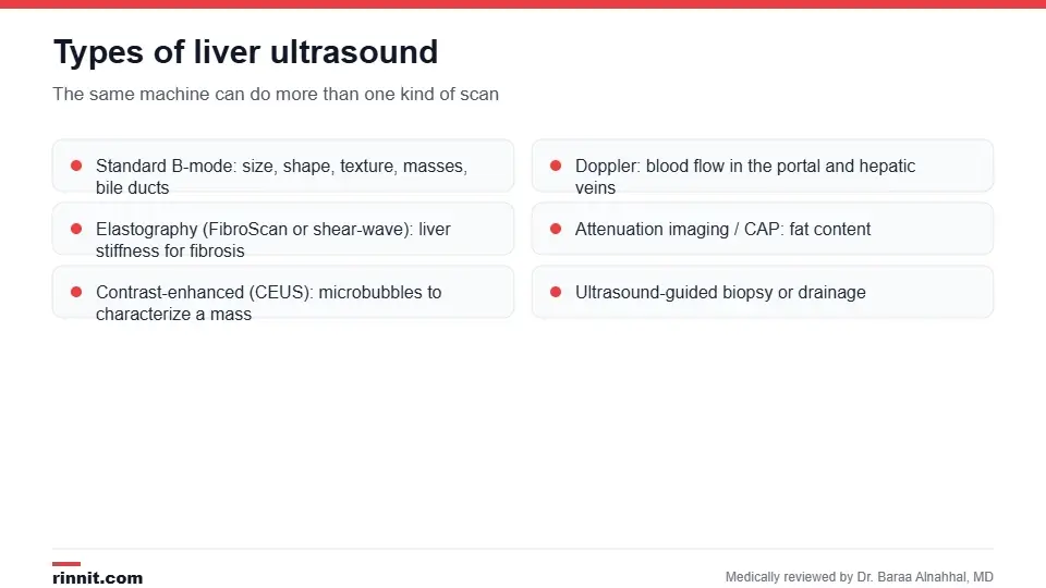
The same machine can perform several kinds of liver scan.
Type | What it adds |
Standard (B-mode) | Size, shape, texture, masses, cysts, bile duct width, fluid in the abdomen |
Doppler | Direction and speed of blood flow in the portal and hepatic veins; detects clots and portal hypertension |
Liver stiffness as a measure of fibrosis and cirrhosis | |
Attenuation imaging / CAP | Amount of fat in the liver |
Contrast-enhanced (CEUS) | Microbubble contrast to characterize a mass as benign or malignant |
Ultrasound-guided procedures | Biopsy of a lesion or drainage of fluid |
Sources: [1]
Why is it done?
Your provider may order a liver ultrasound for abnormal liver blood tests, pain in the right upper abdomen, jaundice, unexplained weight loss, or to monitor a known condition. It can detect or help evaluate: Sources: [1]
Fatty liver disease (MASLD) and its severity.
Hepatitis and cirrhosis.
Cysts, hemangiomas, adenomas and liver cancer, including surveillance in people with cirrhosis or hepatitis B.
Gallstones, cholecystitis and stones in the bile duct (choledocholithiasis).
Portal vein thrombosis, Budd-Chiari syndrome and portal hypertension.
Fluid in the abdomen (ascites).
How do I prepare?
Do not eat for 6–12 hours before the scan; water is usually allowed.
Take regular medicines with a small sip of water unless told otherwise.
Wear loose clothing; you will lift your top or change into a gown.
Tell the staff if you are pregnant or have had recent barium tests, which can interfere with the images. Sources: [1]
What happens during the scan?
You lie on your back, then on your left side, on an exam table.
Warm gel is spread over the right upper abdomen.
The sonographer presses the transducer against the skin and moves it to view the liver from several angles.
You will be asked to take deep breaths and hold them so the liver moves down from under the ribs.
Doppler or elastography measurements are added if ordered.
The gel is wiped off and you can eat and drive as usual.
The scan takes about 30 minutes, up to an hour if several techniques are combined. It is painless, though pressure over a tender area can be uncomfortable. Sources: [1]
Understanding your results
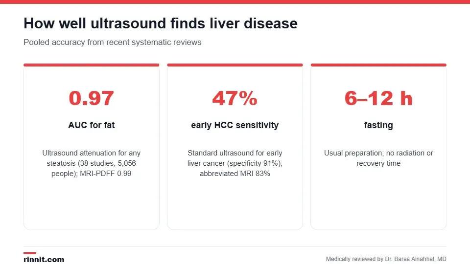
How well ultrasound performs depends on the question being asked.
Finding | What it may mean |
Bright (echogenic) liver | Fat in the liver (steatosis) |
Coarse, nodular texture with an enlarged spleen | Cirrhosis; Doppler may show reversed portal flow |
Simple cyst | Benign; usually no follow-up |
Hemangioma | Benign tangle of vessels; may be confirmed with CEUS or MRI |
Solid mass in a cirrhotic liver | Needs contrast CT or MRI to rule out hepatocellular carcinoma |
Dilated bile ducts | Blockage by a stone, stricture or tumor |
Thickened gallbladder wall, stones, fluid | Cholecystitis |
A radiologist writes the report within a few days. Ultrasound is highly accurate for measuring liver fat (AUC 0.97 for any steatosis, close to MRI), but it detects only about half of early liver cancers in surveillance, so people at high risk may be offered MRI in addition. Sources: [1, 2, 3]
Risks and limitations
There are no side effects or radiation. Image quality can be limited by bowel gas, obesity and the position of the liver under the ribs, and small or deep lesions may be missed. Contrast microbubbles rarely cause allergic reactions. Sources: [1]
Conclusion
A liver ultrasound is the first-line, no-radiation way to look at your liver, and with elastography and attenuation imaging it can now grade scarring and fat without a biopsy. Know its limits too: for cancer surveillance in a cirrhotic liver, ask your provider whether MRI should be added.
Keep learning: see how elastography stages fibrosis and what an abdominal ultrasound covers.
Frequently asked questions
Why do I need to fast before a liver ultrasound?
Fasting reduces bowel gas, which blocks sound waves, and lets the gallbladder fill so stones and wall thickening are easier to see.
How long does a liver ultrasound take?
About 30 minutes; up to an hour if Doppler, elastography or contrast is added.
Does it hurt?
No. You may feel pressure from the probe, especially over a tender area, but there is no pain or radiation.
Can ultrasound diagnose fatty liver?
Yes. Ultrasound attenuation imaging detects any liver fat with an AUC of 0.97, almost as accurate as MRI.
Can ultrasound detect liver cancer?
It can, but it detects only about 47% of early hepatocellular carcinomas in surveillance studies. MRI is more sensitive for high-risk patients.
What does a "bright" or echogenic liver mean?
It usually indicates fat in the liver. Your provider may order elastography or blood tests to assess scarring.
What is a liver hemangioma?
A benign cluster of blood vessels, common and usually harmless. It may be confirmed with contrast ultrasound or MRI.
When will I get my results?
A radiologist's report is usually ready within a few days and reviewed with you by your provider.
What is the difference between ultrasound and elastography?
Elastography is an ultrasound technique that measures liver stiffness to estimate scarring; a standard ultrasound shows structure but not stiffness.
What happens if a mass is found?
Simple cysts need no follow-up. Solid masses are usually evaluated with contrast CT or MRI, and sometimes a biopsy.
Related Reading
Liver-Spleen Scan: Nuclear Imaging of Liver and Spleen Function explains the nuclear scan that shows liver and spleen function.
Antimitochondrial Antibody (AMA) Test for Primary Biliary Cholangitis explains the blood test that helps diagnose primary biliary cholangitis.
Contrast-Enhanced Ultrasound (CEUS): Uses, Safety and What to Expect explains the microbubble ultrasound that shows blood flow without radiation.
Elastography: Types, Preparation, What to Expect and Liver Fibrosis Stages the ultrasound-based stiffness test used to stage liver fibrosis.
Abdominal Ultrasound: Why It's Done, How to Prepare, What to Expect and Results the broader scan that includes the liver, gallbladder and kidneys.
Fatty Liver Disease (MASLD): The Silent Liver Condition Linked to Weight, Diabetes, and Metabolic Health — Causes, Testing, and Treatment the most common finding on liver ultrasound.
Cirrhosis of the Liver: What It Is, How Permanent Scarring Damages Your Liver, and What Treatment Involves what a nodular liver on ultrasound means.
Liver Cancer: A Comprehensive Guide to Symptoms, Causes, and Treatment why people with cirrhosis have surveillance ultrasounds.
References
Cleveland Clinic. Liver Ultrasound: Why It's Done, Prep, Procedure & Results. Cleveland Clinic. Last updated November 21, 2023. https://my.clevelandclinic.org/health/diagnostics/liver-ultrasound
Wu S, Pan J, Song M, et al. Performance of magnetic resonance imaging and ultrasound for identifying the different degrees of hepatic steatosis: a systematic review and meta-analysis. Academic Radiology. 2025;32(11):6528-6540. PMID 40164534. https://doi.org/10.1016/j.acra.2025.03.008
Liu J, Zhong P, Ma W, et al. Comparison of abbreviated MRI and ultrasonography in the detection of early hepatocellular carcinoma: a meta-analysis. Clinical and Translational Oncology. 2026;28(11):5355-5368. PMID 42219433. https://doi.org/10.1007/s12094-026-04431-4
Medical disclaimer
This article is for general education only and does not replace advice from a qualified healthcare professional. Talk to your doctor about your own diagnosis and treatment, and seek emergency care right away for severe or sudden symptoms.



Comments