Contrast-Enhanced Ultrasound (CEUS): Uses, Safety and What to Expect

Updated: 3 days ago
Written and medically reviewed by Dr. Baraa Alnahhal, MD · Last reviewed: November 2026 · How we review
Quick Answer
Contrast-enhanced ultrasound (CEUS) is an ultrasound exam that uses an IV contrast agent made of tiny gas-filled microbubbles to show blood flow in organs and blood vessels in real time. It's most often used to tell benign from cancerous liver lesions. The contrast isn't harmful to the kidneys and the test uses no radiation. Serious reactions are rare.
What the research says
CEUS accurately characterizes liver lesions: in a German multicenter study of more than 1,300 focal liver lesions, CEUS reached an overall diagnostic accuracy of 90.3%, with 95.8% sensitivity for malignant lesions and 83.1% specificity for benign ones (Strobel D et al., Ultraschall in der Medizin, 2008) [2]
CEUS performed as well as CT: a prospective multicenter comparison in routine practice found CEUS was equivalent to CT for telling apart liver tumors, and the authors recommend CEUS before CT to avoid radiation and unnecessary biopsies in many cases (Seitz K et al., Ultraschall in der Medizin, 2009) [3]
Key takeaways
CEUS uses microbubble contrast to show blood flow on ultrasound.
It's commonly used to characterize liver and kidney lesions.
The contrast is safe for the kidneys, and there's no radiation.
Mild side effects affect about 5% of people; anaphylaxis is extremely rare.
You can go home right after the test.
What is CEUS?
A standard ultrasound uses sound waves to make pictures. In CEUS, the microbubbles in the contrast are smaller than red blood cells. They vibrate and strongly reflect sound waves, making areas with blood flow look bright. Because cancer and other diseased tissue often have more blood flow, they tend to light up differently from normal tissue. The gas dissolves in your blood and is breathed out within minutes. Sources: [1]
Why it's done

CEUS shows how lesions take up blood flow.
Telling apart benign and cancerous liver lesions, such as liver hemangiomas, focal nodular hyperplasia, liver cancer and metastases.
Checking kidney masses, kidney cysts and kidney infections.
Monitoring how tumors respond to treatment.
Looking at the bladder, blood vessels, bowel, gallbladder, heart, pancreas and spleen. Sources: [1, 2]
CEUS vs. CT and MRI
Feature | CEUS | CT or MRI with contrast |
Radiation | None | CT uses X-rays; MRI doesn't |
Kidney safety of contrast | Not harmful to the kidneys | CT contrast can affect the kidneys |
Real-time imaging | Yes | No |
Best for | Liver, kidney and targeted questions | Wider views of the body |
CEUS can be used alongside CT or MRI, or sometimes in their place. Sources: [1, 3]
How to prepare
Wear loose, comfortable clothing, or change into a gown.
Some exams require fasting or a full bladder; follow your provider's instructions.
Tell your provider about allergies and heart or lung conditions. Sources: [1]
What happens during the test
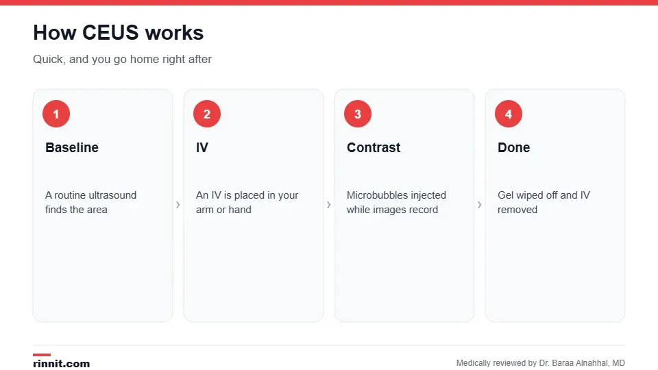
The test is quick and you can leave right away.
A routine ultrasound is done first with gel and a handheld transducer to find the area of interest.
An IV is placed in your arm or hand.
The contrast is injected, and images are recorded as it flows through the tissue; this may be repeated.
The gel is wiped off, the IV is removed and you can go home. Sources: [1]
A radiologist interprets the images and sends results to your provider. Sources: [1]
Risks and side effects
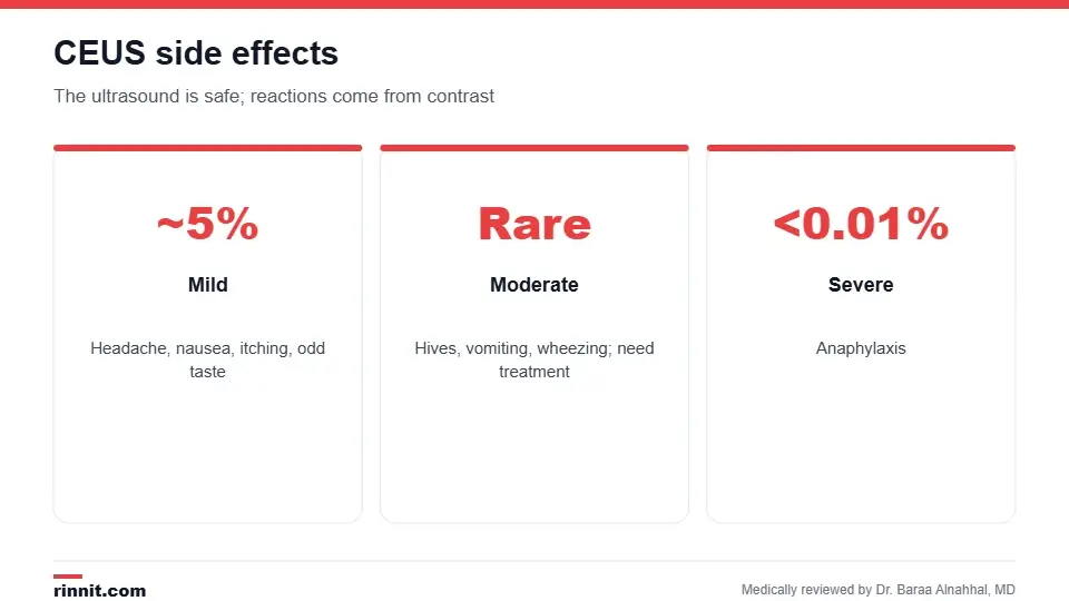
The ultrasound itself is safe; side effects come from the contrast.
Type | Examples | How common |
Mild | Headache, nausea, itching or hives, warmth at the IV site, odd taste | About 5% |
Moderate | Severe hives, vomiting, wheezing, facial swelling, fainting | Uncommon; need treatment |
Severe | Anaphylaxis | Less than 0.01% |
Sources: [1]
Conclusion
CEUS adds real-time blood flow information to a regular ultrasound without radiation or kidney risk. It's a valuable, safe tool for characterizing liver and kidney lesions and can sometimes spare you a CT scan or biopsy.
Keep learning: read about the liver ultrasound and kidney ultrasound.
Frequently asked questions
What is contrast-enhanced ultrasound?
An ultrasound that uses microbubble contrast to show blood flow.
What is CEUS used for?
Most often to characterize liver and kidney lesions.
Is CEUS contrast safe for the kidneys?
Yes. It isn't harmful to the kidneys.
Does CEUS use radiation?
No.
What are microbubbles?
Tiny gas-filled bubbles that reflect sound waves strongly.
What side effects can happen?
Mild headache, nausea or itching in about 5% of people.
Are serious reactions common?
No. Anaphylaxis occurs in less than 0.01% of people.
Do I need to fast?
Sometimes; follow your provider's instructions.
Can children have CEUS?
Yes. It may be safer than CT because it avoids X-rays.
Can I go home after the test?
Yes, right away.
Related Reading
Liver Ultrasound: Why It's Done, How to Prepare, Types and What Results Mean the standard ultrasound of the liver.
Kidney Ultrasound: Why It's Done, How to Prepare and What It Shows ultrasound of the kidneys.
Abdominal Ultrasound: Why It's Done, How to Prepare, What to Expect and Results ultrasound of the belly organs.
Liver Lesions: A Guide to Benign and Malignant Growths the lesions CEUS most often helps characterize.
Gadolinium Contrast for MRI: How It Works, Side Effects and Kidney Safety the contrast used for MRI.
References
Cleveland Clinic. Contrast-Enhanced Ultrasound (CEUS): What It Is & Procedure. Cleveland Clinic. Last updated April 15, 2022. https://my.clevelandclinic.org/health/diagnostics/22754-contrast-enhanced-ultrasound-ceus
Strobel D, Seitz K, Blank W, et al. Contrast-enhanced ultrasound for the characterization of focal liver lesions: diagnostic accuracy in clinical practice (DEGUM multicenter trial). Ultraschall in der Medizin. 2008;29(5):499-505. PMID 19241506. https://doi.org/10.1055/s-2008-1027806
Seitz K, Strobel D, Bernatik T, et al. Contrast-enhanced ultrasound (CEUS) for the characterization of focal liver lesions: prospective comparison in clinical practice: CEUS vs. CT (DEGUM multicenter trial). Ultraschall in der Medizin. 2009;30(4):383-389. PMID 19688670. https://doi.org/10.1055/s-0028-1109673
Medical disclaimer
This article is for general education only and does not replace advice from a qualified healthcare professional. Talk to your doctor about your own diagnosis and treatment, and seek emergency care right away for severe or sudden symptoms.



Comments