Elbow X-Ray: What It Shows, Views Taken and What to Expect

Updated: 3 days ago
Written and medically reviewed by Dr. Baraa Alnahhal, MD · Last reviewed: November 2026 · How we review
Quick Answer
An elbow X-ray uses a small amount of radiation to make pictures of the bones of the elbow joint. It's often the first test after an elbow injury or for pain, swelling or deformity. It can show fractures, dislocations and arthritis. Tendon problems such as tennis elbow don't show on X-ray and are usually diagnosed by exam.
What the research says
A simple bedside test helps decide who needs an X-ray: in 2,127 adults and children with elbow injuries, being unable to fully straighten the elbow detected fractures with 96.8% sensitivity; people who could fully extend their elbow had only about a 1.6% (adults) or 4.2% (children) chance of fracture, and those who couldn't had nearly a 50% chance (Appelboam A et al., BMJ, 2008) [2]
A hidden fracture can show up as a "fat pad sign": in 45 children whose elbow X-rays showed only an elevated posterior fat pad after injury, 76% were later found to have a fracture, so these children are usually treated as if they have a nondisplaced fracture (Skaggs DL et al., Journal of Bone and Joint Surgery, 1999) [3]
Key takeaways
Shows the elbow bones to find fractures, dislocations, arthritis, cysts, infection and tumors.
At least three views: front, side and angled.
Takes about 15 minutes; remove jewelry and tell staff if you might be pregnant.
Low radiation; children get lower doses.
Doesn't show tendons, so tennis and golfer's elbow are diagnosed by exam.
Why is it done?
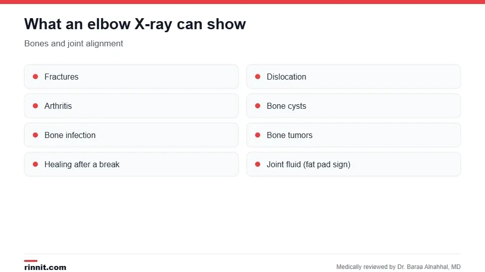
An elbow X-ray shows bones and joint alignment.
Pain, swelling, tenderness or deformity after a fall or injury.
To check for a fracture or a dislocated elbow.
To look for arthritis, bursitis, bone cysts, bone infection (osteomyelitis) or bone tumors.
To check that a broken bone is lined up and healing.
Before and after elbow surgery or elbow replacement. Sources: [1]
Views taken
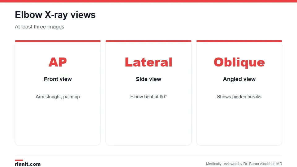
A standard elbow X-ray includes at least three views.
View | Position | What it shows best |
Anteroposterior (AP) | Arm straight, palm up | Front view of the joint |
Lateral | Elbow bent at 90 degrees | Side view, joint fluid and fat pads |
Oblique | Arm angled | Small or hidden fractures |
Sources: [1, 3]
How do I prepare?
Wear comfortable clothing with short or loose sleeves.
Remove jewelry, watches and other metal.
Tell the technologist if you are or might be pregnant. Sources: [1]
What happens during the X-ray?
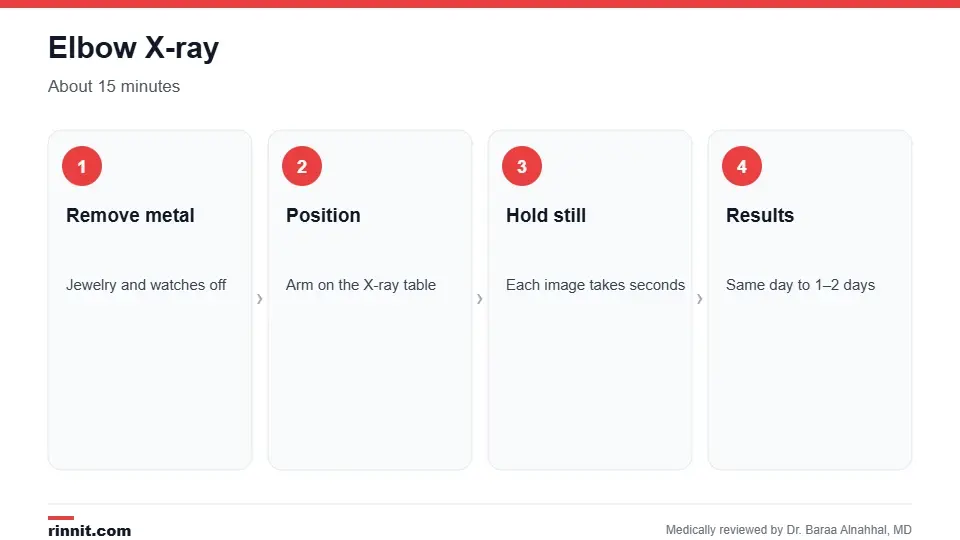
An elbow X-ray takes about 15 minutes.
You sit next to the X-ray table and rest your arm on it.
The technologist positions your arm, sometimes with foam pads to hold it still, and may place a lead apron over your body.
They step behind a screen while each image is taken; you hold still for a few seconds.
They reposition your arm for each view. Sources: [1]
If moving your arm hurts, tell the technologist; they can adjust positions. The exam usually takes about 15 minutes or a bit longer. Sources: [1]
Understanding your results
A radiologist reviews the images. In an emergency, results may be available almost right away; otherwise, they usually reach your provider within one to two days. Besides obvious breaks, the radiologist looks for subtle signs such as a raised fat pad, which can point to blood or fluid in the joint from a hidden fracture, especially in children. If the X-ray is unclear, your provider may order more X-rays, a CT or an MRI. Sources: [1, 3]
Because X-rays don't show tendons well, tennis elbow and golfer's elbow are usually diagnosed with a physical exam; an X-ray may still be done to rule out a fracture or arthritis. Sources: [1]
Risks
The radiation dose is small. Precautions are taken during pregnancy, and children are given lower doses. Sources: [1]
Conclusion
An elbow X-ray is a fast, simple way to check the bones of your elbow after an injury or when pain won't go away. Most results are back within a day or two, and they guide whether you need a cast, a sling, more imaging or surgery.
Keep learning: read about the general X-ray and the wrist X-ray.
Frequently asked questions
What does an elbow X-ray show?
The bones of the elbow, including fractures, dislocations and arthritis.
How many views are taken?
At least three: front, side and angled.
How long does it take?
About 15 minutes.
Do I need to prepare?
Just remove jewelry and wear loose sleeves.
Is the radiation safe?
Yes, the dose is small; tell staff if you might be pregnant.
Can an X-ray show tennis elbow?
No. Tendon problems are diagnosed by exam; an X-ray rules out other causes.
What is a fat pad sign?
A sign on X-ray that can point to a hidden fracture.
Will an X-ray hurt my injured elbow?
It's painless, though positioning a sore arm can be uncomfortable.
When will I get results?
Right away in emergencies; otherwise within one to two days.
What if the X-ray is normal but I still hurt?
Your provider may order more imaging or a follow-up X-ray.
Related Reading
Dislocated Elbow: An Injury That Needs the Emergency Room — What Happens, the 3 Severity Types, Symptoms, Treatment and Recovery Time — What You Need to Know a joint injury diagnosed with X-ray.
Tennis Elbow: Symptoms, Causes, and Treatment outer elbow pain diagnosed by exam.
Golfer's Elbow: Why the Inside of Your Elbow Hurts, and What Actually Heals It inner elbow pain from tendon strain.
Elbow Surgery: Types, Procedure, Risks and Recovery operations that X-rays help plan.
Elbow Replacement Surgery: A Comprehensive Guide to Arthroplasty joint replacement for severe elbow damage.
References
Cleveland Clinic. Elbow X-Ray Anatomy, Procedure & What to Expect. Cleveland Clinic. Last updated July 18, 2022. https://my.clevelandclinic.org/health/diagnostics/23507-elbow-x-ray
Appelboam A, Reuben AD, Benger JR, et al. Elbow extension test to rule out elbow fracture: multicentre, prospective validation and observational study of diagnostic accuracy in adults and children. BMJ. 2008;337:a2428. PMID 19066257. https://doi.org/10.1136/bmj.a2428
Skaggs DL, Mirzayan R. The posterior fat pad sign in association with occult fracture of the elbow in children. J Bone Joint Surg Am. 1999;81(10):1429-1433. PMID 10535592. https://doi.org/10.2106/00004623-199910000-00007
Medical disclaimer
This article is for general education only and does not replace advice from a qualified healthcare professional. Talk to your doctor about your own diagnosis and treatment, and seek emergency care right away for severe or sudden symptoms.



Comments