Understanding the Brachiocephalic Vein: Anatomy, Function, and Health

Updated: 3 days ago
Medically reviewed by Dr. Baraa Alnahhal, MD · Last reviewed: October 2026
TL;DR: The brachiocephalic vein, also called the innominate vein, is a paired vein in the upper chest that returns oxygen-poor blood from the head, neck, and arms to the heart. Each one forms where the subclavian and internal jugular veins meet, and the right and left veins join to form the superior vena cava. [1] Unlike many veins, they have no valves. [1] They are often used for central venous catheters, which can lead to clots or narrowing.
Get urgent care: Sudden swelling of the face, neck, or arm, trouble breathing, or chest pain, especially if you have a central line, dialysis access, or a chest tumor, can signal a blood clot or a blocked vein. Call your local emergency number.
Quick Answer: What are the brachiocephalic veins?
The brachiocephalic veins are two major veins of the upper body, one on each side, that collect oxygen-poor blood from the head, neck and arms. [1] They join to form the superior vena cava, which empties into the heart's right atrium. Unlike many veins, they have no valves, and providers often use them for central venous access. [1]
What the research says
A retroaortic left brachiocephalic vein is uncommon but linked to heart defects. In a review of 5,218 children with congenital heart disease, the variation was found in 27 (0.5%), and 25 of those 27 (93%) had tetralogy of Fallot (Curtil et al., Surg Radiol Anat, 1999) [2].
Catheter width affects clot risk in the arm veins. In a meta-analysis of 40 studies, symptomatic deep vein thrombosis occurred in 0.89%, 3.26%, 5.46%, and 10.66% of patients with 3, 4, 5, and 6 French peripherally inserted central catheters (Bahl et al., Clin Appl Thromb Hemost, 2023) [3].
Catheter-related clots in people with cancer usually respond well to treatment. Across 29 studies and 2,836 patients, the pooled 3-month rate of recurrent clots was 0.56% and the pooled rate of major bleeding was 2.34% (Wang et al., J Thromb Haemost, 2024) [4].
Key takeaways
The brachiocephalic vein, also called the innominate vein, is paired: you have a right and a left.
Each forms where the subclavian vein and the internal jugular vein meet, deep in the upper chest below the collarbone.
The right vein is about 2 to 3 cm long and runs almost straight down. The left is about 6 cm long and crosses the chest to join it. [1]
They have no valves, which makes them useful for central venous access but also exposes them to catheter-related problems.
Stenosis, blood clots, perforation, and compression from chest tumors can affect them. Swelling in the neck, face, or arms deserves a medical check.
What does the brachiocephalic vein do?
The main job of the brachiocephalic veins is to return oxygen-poor blood from the head, neck, and arms to the heart. [1] They are short segments of a much larger venous system, but they are the final collection points before blood reaches the superior vena cava. From there, blood enters the right atrium, moves through the right ventricle, and travels to the lungs to pick up oxygen.
If these veins are narrowed or blocked, drainage from the upper body can back up. That is why swelling in the neck, face, or arms is an important warning sign.
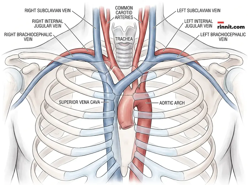
What is the structure and path of the brachiocephalic vein?
The brachiocephalic veins are deep veins in the upper chest, just below the collarbones. Each one forms where the internal jugular vein and the subclavian vein join. [1] The two sides share a name and a job, but they differ in length and path:
Feature | Right brachiocephalic vein | Left brachiocephalic vein |
Length | About 2 to 3 centimeters | About 6 centimeters |
Orientation | Travels almost vertically down toward the heart | Travels horizontally at a slight downward angle |
Path | Direct, short descent | Passes above the aortic arch and in front of the carotid arteries [1] |
Union | Joins the left vein to form the superior vena cava | Joins the right vein to form the superior vena cava |
Like other veins, the wall has three layers: the tunica adventitia (outer layer) gives structure and shape, the tunica media (middle layer) has smooth muscle that lets the vein widen or narrow, and the tunica intima (inner layer) is a smooth lining that lets blood flow easily. Unlike many veins, the brachiocephalic veins have no valves. [1]
Many veins drain into the brachiocephalic veins, including the:
Internal jugular veins
Subclavian veins
Inferior thyroid veins
Internal thoracic veins (also called internal mammary veins), covered in our guide to the internal thoracic vein
Vertebral veins
Left superior intercostal vein [1]

Why is the brachiocephalic vein clinically significant, and how does it vary?
Providers often use the brachiocephalic veins for central venous access, inserting a thin, flexible tube to deliver fluids or medicines directly into the circulation. Complications can include a hole in the vein (perforation) or deep vein thrombosis (DVT) in the upper body, and, rarely, a pseudoaneurysm. [1] For more on catheters, see our guides to PICC lines and non-tunneled central venous catheters. In people on dialysis, an arteriovenous fistula often involves this vein, and vein narrowing (stenosis) can be a complication [1] (see Dialysis Fistula (AV Fistula)).
Some people are born with anatomical variations. The most common is a retroaortic brachiocephalic vein [1], in which the left vein passes behind the ascending aorta instead of in front of it. Variations affect about 1 in 250 babies. [1] On their own they are usually harmless, but they often occur alongside congenital heart disease, so providers may order imaging before surgeries or procedures such as connecting to a heart-lung machine or placing a pacemaker.
Condition | Common cause | Impact on health |
Vein stenosis (narrowing) | Medical procedures or dialysis access | Restricted blood flow and higher pressure behind the narrowing |
Deep vein thrombosis | Catheter complications or slowed flow | Blood clots and a risk of pulmonary embolism |
Perforation | Catheter placement | Bleeding and damage to the vein |
Compression | Mediastinal tumors or cancer spread to the chest [1] | Blocked return of blood from the upper body |
To check these veins, providers may use duplex ultrasound, computed tomography (CT), magnetic resonance imaging (MRI), or a venogram (an X-ray of the veins after dye is injected). [1]

How can you keep your veins, including the brachiocephalic vein, healthy?
A heart-healthy lifestyle keeps the brachiocephalic veins and other blood vessels working well: avoid tobacco, drink plenty of water, eat well, walk regularly and take movement breaks.
Avoid smoking and all tobacco products.
Drink plenty of water.
Eat a heart-healthy diet, which can help lower blood pressure, cholesterol, and body weight.
Walk for 30 minutes at least five days a week [1], after checking with your provider before starting a new routine.
Take short movement breaks during the day to keep blood moving.
When should you see a healthcare provider about your brachiocephalic vein?
Talk to a provider if you notice unusual swelling in your neck, face, or arms, especially if you have a central line, dialysis access, or a known chest tumor. Early checks matter because blockages in these veins can be treated best when found early. Seek emergency care for sudden swelling with trouble breathing or chest pain.
Conclusion
The brachiocephalic veins are the bridge between the veins of your head, neck, and arms and the heart. Their lack of valves and their role in forming the superior vena cava make them both essential and clinically important. A heart-healthy lifestyle and attention to warning signs help protect this key part of your circulation.
Related reading
Internal Thoracic Vein: Anatomy, Function, and Surgical Role
Understanding PICC Lines: A Guide to Peripherally Inserted Central Catheters
Understanding Non-Tunneled Central Venous Catheters: A Guide to Short-Term Vascular Access
Dialysis Fistula (AV Fistula): A Guide to Long-Term Hemodialysis Access
Understanding the Brachial Artery: Anatomy, Function, and Health
Frequently Asked Questions
What is the other name for the brachiocephalic vein?
The brachiocephalic vein is also called the innominate vein.
How many brachiocephalic veins do I have?
Two: a right and a left brachiocephalic vein.
What is the main function of these veins?
The brachiocephalic veins return oxygen-poor blood from the head, neck, and arms to the heart.
Which veins join to form a brachiocephalic vein?
The subclavian vein and the internal jugular vein on the same side.
What large vessel do they form?
The right and left veins merge to form the superior vena cava.
Where are these veins located?
Deep in the upper chest, below the collarbones.
Why is the left vein longer than the right?
The left vein runs horizontally across the chest to reach the right vein, while the right vein drops almost straight down.
Do brachiocephalic veins have valves?
No. Unlike many veins, they do not contain valves.
What are the three layers of the vein wall?
The tunica adventitia (outer), tunica media (middle), and tunica intima (inner).
What is a retroaortic brachiocephalic vein?
A variation in which the left vein passes behind the ascending aorta instead of in front of it.
How common are anatomical variations?
Anatomical variations affect about 1 in 250 babies. Many occur alongside congenital heart disease.
What is vein stenosis?
Vein stenosis is narrowing of the vein, which can follow medical procedures or dialysis access.
How do doctors check these veins?
With duplex ultrasound, CT, MRI, or a venogram.
Can tumors affect the brachiocephalic vein?
Yes. Growths in the chest can press on the vein and block blood flow.
How can I keep my veins healthy?
Do not smoke, drink water, eat a heart-healthy diet, and keep moving through the day.
Sources
Cleveland Clinic. Brachiocephalic Vein. Cleveland Clinic. Last updated July 8, 2022. https://my.clevelandclinic.org/health/body/23466-brachiocephalic-vein
Curtil A, Tronc F, Champsaur G, et al. The left retro-aortic brachiocephalic vein: morphologic data and diagnostic ultrasound in 27 cases. Surg Radiol Anat. 1999;21(4):251-254. PMID 10549081. https://doi.org/10.1007/BF01631395
Bahl A, Alsbrooks K, Gala S, et al. Symptomatic deep vein thrombosis associated with peripherally inserted central catheters of different diameters: a systematic review and meta-analysis. Clin Appl Thromb Hemost. 2023;29:10760296221144041. PMID 37366542. https://doi.org/10.1177/10760296221144041
Wang TF, Kou R, Carrier M, et al. Management of catheter-related upper extremity deep vein thrombosis in patients with cancer: a systematic review and meta-analysis. J Thromb Haemost. 2024;22(3):749-764. PMID 38065528. https://doi.org/10.1016/j.jtha.2023.11.017
Medical disclaimer
The information in this article is for educational purposes only and is not a substitute for professional medical advice, diagnosis, or treatment. Always seek the advice of your physician or another qualified health provider with any questions you may have about a medical condition. Never disregard professional medical advice or delay seeking it because of something you have read here. If you think you may have a medical emergency, call your doctor or your local emergency number immediately.



Comments