Internal Thoracic Vein: Anatomy, Function, and Surgical Role

Updated: 2 days ago
Medically reviewed by Dr. Baraa Alnahhal, MD · Last reviewed: September 2026
TL;DR
The internal thoracic vein is a critical blood vessel located deep within the chest that returns oxygen-poor blood from the chest wall and breast to the heart. Beyond its circulatory role, it is frequently utilized by surgeons as a reliable linking vessel in breast reconstruction procedures.
Quick Answer
The internal thoracic vein, formerly known as the internal mammary vein, is a paired blood vessel running vertically on either side of the breastbone. Its primary function is to drain oxygen-poor blood from the chest wall, breast, diaphragm, and heart sac (pericardium) back to the heart. It is clinically significant due to its predictable anatomy, which makes it an ideal choice for connecting blood vessels during complex reconstructive surgeries.
What Is the Internal Thoracic Vein?
The internal thoracic vein is a specialized blood vessel located deep within the thoracic cavity. It exists as a pair, with one vein positioned on each side of the chest. Historically referred to as the internal mammary vein, this vessel plays a quiet but vital role in the body's circulatory network.
Each internal thoracic vein travels alongside a corresponding internal thoracic artery. While the artery carries oxygen-rich blood away from the heart to supply the chest tissues, the vein performs the opposite task, collecting oxygen-poor blood to begin its journey back to the lungs for re-oxygenation.
Function and Circulatory Role
The primary responsibility of the internal thoracic vein is to support the return of blood to the heart and lungs. By draining various structures within the chest, it ensures that metabolic waste products are removed and that blood can be refreshed with oxygen.
The vein is responsible for draining blood from several key areas:
The Breast: It collects blood from the mammary tissues.
The Diaphragm: It supports the primary muscle used for breathing.
The Pericardium: It drains the protective sac surrounding the heart.
The Chest Wall: It collects blood from the muscles and bones that protect the thoracic organs.
Once the blood enters the internal thoracic vein, it flows upward toward the neck, eventually reaching the heart's upper right chamber.

Anatomy and Pathway
The internal thoracic veins are oriented vertically and run deep within the chest, positioned close to the breastbone (sternum). They span the distance from the upper abdominal region to the collarbone.
The Pathway
The vein begins at the junction of two smaller vessels: the musculophrenic vein and the superior epigastric vein. From this starting point in the upper belly, it travels upward alongside the sternum.
Ultimately, the right internal thoracic vein empties into the right brachiocephalic vein, while the left internal thoracic vein empties into the left brachiocephalic vein. These brachiocephalic veins then merge to form the superior vena cava, the large vessel that delivers blood directly into the heart.
Tributaries
As the internal thoracic vein travels, it receives blood from several "tributaries," which are smaller veins that flow into it. These include:
Anterior Intercostal Veins: These drain the spaces between the ribs at the front of the body.
Pericardiophrenic Veins: These drain structures located in the mediastinum, the space in the middle of the chest.
Feature | Description |
Type | Paired vein (Left and Right) |
Location | Deep chest, lateral to the sternum |
Origin | Junction of musculophrenic and superior epigastric veins |
Termination | Brachiocephalic vein |
Primary Drainage | Breast, diaphragm, pericardium, chest wall |
Anatomical Variations
Individual anatomy can vary, and the internal thoracic vein is no exception. A common variant is bifurcation, where the vein splits into two separate vessels partway through its course. In these cases, two veins will travel upward on either side of the internal thoracic artery. These structural differences are considered normal and do not negatively impact the vein's ability to function.
Clinical and Surgical Significance
While the internal thoracic vein is essential for daily circulation, it gains significant attention in the field of surgery, particularly in breast reconstruction.
Role in Reconstruction
During flap reconstruction surgery, tissue is moved from one part of the body to form a new breast. For this tissue to survive, its blood vessels must be connected to the circulatory system in the chest. Surgeons often choose the internal thoracic vein as the "linking vessel" for this connection. It is preferred because it is centrally located, typically healthy, and follows a predictable anatomical path.
Potential Conditions
The internal thoracic vein can be affected by specific injuries or medical complications:
Chest Trauma: Severe injuries to the chest can damage the vein and surrounding tissues.
Medical Procedures: Procedures such as pericardiocentesis (draining fluid from the heart sac) carry a rare risk of damaging the vessel. To minimize this, healthcare providers use ultrasound imaging to guide their instruments safely.
Clinical Aspect | Details |
Surgical Use | Linking vessel for breast flap reconstruction |
Preferred Traits | Central location, predictable path, vessel health |
Diagnostic Tool | Ultrasound for procedure guidance |
Risk Factors | Chest trauma, rare procedural complications |

Conclusion
The internal thoracic vein is a fundamental component of the thoracic circulatory system. By providing a reliable drainage pathway for the breast, diaphragm, and chest wall, it supports systemic health. Its predictable anatomy further makes it an invaluable asset in modern reconstructive surgery, highlighting its importance in both natural function and clinical intervention.
Proactive Care and Monitoring
Maintaining vascular health involves a combination of healthy lifestyle choices and regular medical checkups. While the internal thoracic vein is located deep within the body, supporting overall circulation benefits all vessels.
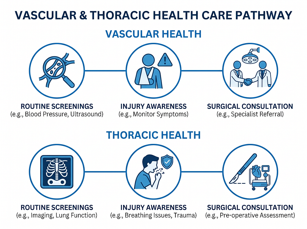
Routine Screenings: Discuss circulatory health with a provider during annual exams.
Injury Awareness: Seek immediate attention for severe chest trauma.
Surgical Consultation: If undergoing reconstruction, discuss the role of chest vessels with your surgical team.
Frequently Asked Questions
1. What is the internal thoracic vein?
It is a paired vein located deep in the chest that returns blood from the chest wall and breast to the heart.
2. What was this vein formerly called?
It was previously known as the internal mammary vein.
3. Where is the internal thoracic vein located?
It runs vertically on both sides of the breastbone (sternum), extending from the upper abdomen to the collarbone.
4. What is the primary function of this vein?
Its main job is to drain oxygen-poor blood from the chest wall, breast, diaphragm, and pericardium.
5. Where does the internal thoracic vein empty?
It empties into the brachiocephalic vein on the corresponding side of the body.
6. What is the superior vena cava's relationship to this vein?
The internal thoracic vein empties into the brachiocephalic vein, which then joins the other brachiocephalic vein to form the superior vena cava.
7. What are the tributaries of the internal thoracic vein?
Primary tributaries include the anterior intercostal veins and the pericardiophrenic veins.
8. What does "bifurcation" mean in relation to this vein?
It refers to a normal anatomical variant where the vein splits into two vessels partway through its course.
9. Why is this vein important in breast reconstruction?
Surgeons use it as a linking vessel to connect the blood supply of transplanted tissue to the chest's circulation.
10. Why do surgeons prefer this vein for surgery?
It is preferred due to its central location, predictable anatomy, and generally healthy condition.
11. Can the internal thoracic vein be damaged?
Yes, it can be harmed by severe chest trauma or, rarely, as a complication of certain medical procedures.
12. How do doctors protect the vein during procedures?
They often use ultrasound imaging to visualize the vessel and guide instruments safely.
13. Does having a bifurcated vein cause health problems?
No, it is a normal anatomical variation that does not affect the vein's function.
14. What structures does the internal thoracic vein drain?
It drains the breast, the diaphragm, the pericardium (heart sac), and the bones and muscles of the chest wall.
15. What artery runs alongside this vein?
The internal thoracic artery runs vertically next to the internal thoracic vein.
Medical Disclaimer: This content is for informational purposes only and does not constitute medical advice. Always seek the guidance of a qualified healthcare professional regarding any medical condition or treatment.
Comments