Understanding Staghorn Calculus: Causes, Symptoms, and Treatments

Updated: 2 days ago
Medically reviewed by Dr. Baraa Alnahhal, MD · Last reviewed: October 2026
Quick Answer: What is a staghorn calculus?
A staghorn calculus is a complex, branching kidney stone that fills the kidney's collecting system like coral or antlers, often made of struvite from chronic urinary tract infections. It can block urine flow, causing kidney swelling and, if untreated, kidney failure. [1] Treatment almost always requires surgery, such as percutaneous nephrolithotomy, to remove the entire stone.
When to get urgent care
Seek emergency care for fever and shaking chills together with flank pain, confusion, a fast heartbeat, or very little urine. These can be signs of a serious infection of the kidney or bloodstream (sepsis) in someone with a kidney stone. For milder symptoms such as ongoing side pain, recurrent urinary infections, or blood in the urine, see a healthcare professional promptly.

What the research says
Most staghorn stones are infection stones. A review explains that most staghorn stones are made of struvite (magnesium ammonium phosphate) and are linked to urinary tract infection by urease-producing bacteria. It identifies the main goals of treatment as becoming stone-free, eradicating infection, and preventing recurrence, with percutaneous nephrolithotomy (PCNL) as the gold-standard operation (Torricelli and Monga, International Braz J Urol, 2020) [2].
Untreated stones carry serious risks. A best-practice review states that staghorn calculi are associated with high morbidity and can lead to recurrent urinary tract infections, urosepsis, kidney deterioration, and death if left untreated, while modern endourology techniques can treat them with minimal morbidity (Sharbaugh et al., Therapeutic Advances in Urology, 2019) [3].
Technique matters for clearing every branch. In a randomized trial of 160 patients (81 vs 79), adding antegrade flexible ureteroscopy during PCNL gave a higher stone-free rate, needed fewer percutaneous tracts, and shortened operating time compared with standard PCNL, with comparable complication rates (Qi et al., Urolithiasis, 2024) [4].
What Is a Staghorn Calculus?
A staghorn calculus is a type of kidney stone with a distinctive branching shape. [1] "Calculus" means stone, and "staghorn" describes how it can look like deer antlers or coral. [1] Unlike typical small kidney stones that can pass through the urinary tract, staghorn stones grow to occupy the renal pelvis and the calyces, the funnel-shaped structures where urine collection begins inside the kidney. To learn about ordinary stones first, see our general guide to kidney stones.
These stones are serious because their growth pattern can cause blockages. If left untreated, the obstruction can make the kidney swell and eventually fail. [1] Staghorn stones are not a common type of kidney stone, and they usually affect one kidney, although both can be involved. [1]
What are the symptoms and warning signs of staghorn calculus?
Symptoms vary. Sometimes the pain from these large stones is surprisingly mild, felt as a dull ache in the side between the ribs and the hip. The infections and complications that come with the stone are often more noticeable.
Persistent side pain: a mild but constant discomfort in the flank.
Fever and chills: signs of an infection trapped by the stone.
Urinary changes: blood (hematuria) or pus (pyuria) in the urine [1], or cloudy urine.
Fatigue: tiredness or feeling generally unwell, often linked to chronic infection.
Symptom type | Common signs |
|---|---|
Pain | Mild to severe ache in the flank (between the ribs and the hip) |
Urinary signs | Blood in the urine, pus in the urine, or cloudy urine |
Infection signs | Fever, shaking chills, and recurrent urinary tract infections |
General health | Fatigue and feeling unwell |
What causes staghorn calculus and who is at risk?
Most staghorn calculi are linked to repeated urinary tract infections (UTIs). Certain bacteria, such as Proteus mirabilis, make an enzyme called urease. [1] It makes urine less acidic and creates conditions in which crystals of magnesium, phosphate, and ammonium (struvite) can form and grow into large, branching stones. [1] See our guides to urinary tract infections and recurrent UTIs.
Other factors can raise the likelihood of stone formation. Differences in the urinary tract anatomy or existing blockages can trap urine and encourage crystals to grow. People who need long-term urinary catheters (see our guide to the Foley catheter) or who have a neurogenic bladder are at higher risk. [1] These stones are more common in women, and in people with spinal cord injuries, Crohn's disease, or certain types of weight-loss surgery. [1]
What are the possible complications of staghorn calculus?
The main danger is that the stone blocks urine flow from the kidney. As the stone fills the renal pelvis, urine backs up, and the kidney and its calyces swell, a condition called hydronephrosis (or caliectasis).
The stone's rough surface can also shelter bacteria. This makes infections hard to clear, because bacteria can hide inside the stone and come back even after antibiotics. If infection reaches the bloodstream, it can cause urosepsis, a life-threatening whole-body response; see our guide to sepsis. Chronic blockage and repeated infection can lead to permanent kidney damage and kidney failure; for sudden loss of kidney function, see acute kidney injury.

How is staghorn calculus diagnosed?
Staghorn calculus diagnosis starts with a medical history and physical exam, then CT imaging, urine tests and blood tests confirm the stone, its size and kidney function. Providers look for a history of recurrent infections and typical flank pain, then use tests to confirm the stone and its size:
Imaging: computed tomography (CT) is considered the most accurate way to show the stone's branching structure. A renal scan may be used to assess how well the kidney works.
Laboratory tests: urinalysis and urine culture can detect bacteria, blood, or pus, and blood tests help evaluate kidney function and signs of systemic infection.
How is staghorn calculus managed and treated?
Treatment almost always involves surgical removal. Because the stone is a home for bacteria, leaving fragments behind can lead to new stones or a return of infection, so the goal is to clear all of the stone material. [1]
Treatment | Description | Main use |
|---|---|---|
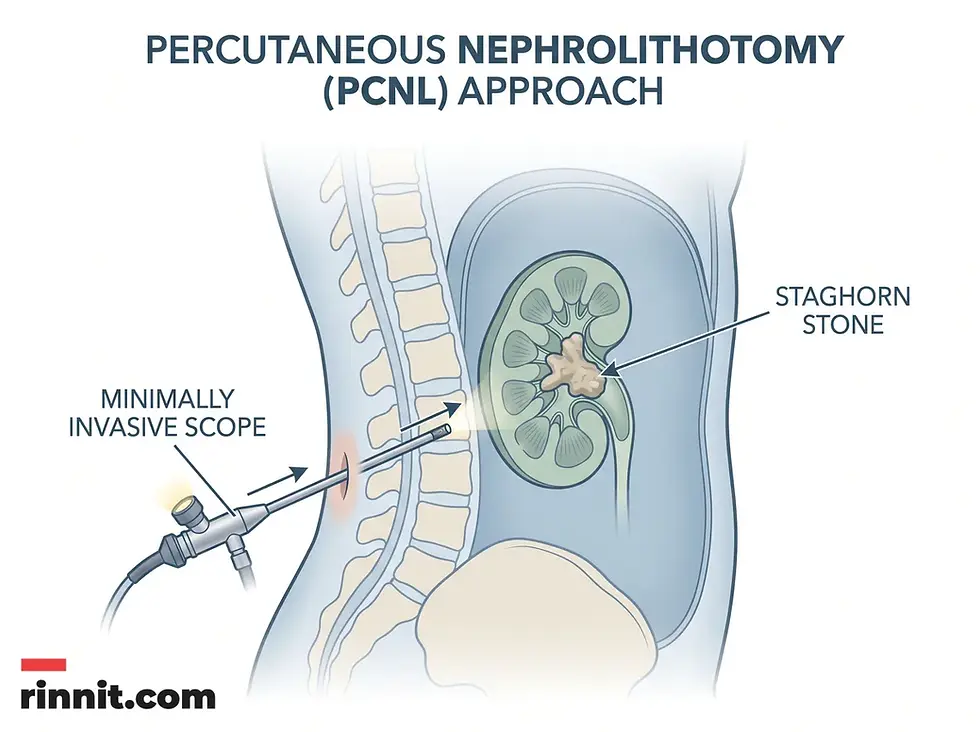
Percutaneous nephrolithotomy (PCNL) | Large stones are broken up and removed through a small incision in the back | First-choice treatment for most staghorn stones |
Ureteroscopy | A scope passed through the ureter to find and remove stone fragments | Smaller fragments, or combined with other methods |
Laser lithotripsy | A laser breaks stones into smaller pieces | Often used during PCNL or ureteroscopy; see our guide to laser lithotripsy |
Ureteral stent | A temporary plastic tube in the ureter to keep urine draining | Relieving a blockage or aiding healing after surgery |
Nephrectomy | Surgical removal of the kidney | Only if the kidney no longer works and is severely damaged |
Learn more about how stones are broken up in our article on laser lithotripsy. Doctors also check urine cultures before surgery so that the right antibiotics are used, and short or longer antibiotic courses and follow-up urine cultures are commonly part of care. [2]
What is recovery and the long-term outlook for staghorn calculus?
Recovery varies with the procedure. Minimally invasive treatments typically need about two to four weeks for full recovery, and larger operations can take longer. Some people go home the same day and others stay a night or two, and you will get specific instructions about activity and heavy lifting.
The outlook is generally good when the stone is completely removed. Afterward, most people can urinate normally and have relief from chronic pain and infections. Follow-up with imaging, urine cultures, and blood tests is usually recommended to catch new stones early, especially if you have underlying risk factors.
How can staghorn calculus be prevented?
Preventing a return means addressing what caused the stone. Staying well hydrated is the most important step because it dilutes the substances that form stones. Depending on the stone's makeup, your provider may suggest dietary changes. If you are prone to chronic infections, your provider may recommend preventive antibiotics or other measures to keep the urine free of stone-forming bacteria, and prompt treatment of every UTI matters.
Conclusion
A staghorn calculus is a serious condition that needs active management. By filling the kidney's internal drainage structures, these branching stones threaten kidney health and overall well-being. Early diagnosis with imaging and complete surgical removal are the keys to preventing permanent kidney damage and restoring normal urinary function.
What are the next steps if you have staghorn calculus?
If you have persistent flank pain, recurrent urinary tract infections, or changes in your urine, see a healthcare professional promptly. Early evaluation and treatment protect your kidneys.
Trusted resources: Cleveland Clinic: Staghorn Calculus, American Urological Association: Staghorn Calculi guideline
Frequently Asked Questions
Is a staghorn calculus the same as a regular kidney stone?
A staghorn calculus is a type of kidney stone, but it is distinguished by its large size and branching shape that fills the kidney's drainage system.
Can I pass a staghorn stone naturally?
No. Staghorn stones are too large to pass through the ureters and almost always need surgery.
Are staghorn stones always painful?
No. The pain is often mild or dull, though it can become severe if there is a complete blockage.
What is the most common cause of these stones?
Recurrent urinary tract infections caused by bacteria that make urease are the main cause.
Who is most at risk for staghorn stones?
Women and people with spinal cord injuries, chronic UTIs, long-term catheters, neurogenic bladder, or certain intestinal conditions or surgeries are at higher risk.
What happens if a staghorn stone is left untreated?
An untreated staghorn stone can lead to chronic infection, severe kidney swelling, sepsis, and permanent kidney failure.
How do doctors find these stones?
Usually with a CT scan, which shows the branching structure within the kidney, along with urine and blood tests.
What is PCNL surgery?
Percutaneous nephrolithotomy is a minimally invasive procedure in which a surgeon removes the stone through a small incision in the back.
Can these stones affect both kidneys?
Usually only one kidney is affected, but both can develop staghorn stones.
Is kidney removal necessary?
Nephrectomy is only considered if the kidney is so severely damaged that it no longer works.
What are struvite stones?
Struvite is a mineral made of magnesium, ammonium, and phosphate, often found in stones caused by infection.
Can diet help prevent staghorn stones?
Staying hydrated and following dietary advice from your provider can help, but treating and preventing infections is just as important.
How long is the recovery after surgery?
Most people recover in about two to four weeks, depending on the procedure's complexity.
Can children get staghorn calculi?
Yes, although it is less common. A review in children found that PCNL is recommended as the first-line surgical treatment, as it is in adults.
Can the stones come back after surgery?
Yes. If the underlying cause, such as infection or poor urine drainage, is not addressed, new stones can form.
Related Reading
Kidney Stones: Symptoms, Causes, and Treatment Guide explains how kidney stones form and are treated.
Percutaneous Nephrolithotomy (PCNL): Procedure, Risks, and Recovery describes the keyhole surgery most often used to remove staghorn stones.
Urinary Tract Infection (UTI): Symptoms, Causes, Treatment, and Prevention covers the urinary infections that can lead to struvite stones.
Extracorporeal Shock Wave Lithotripsy (ESWL): Procedure, Recovery, Risks and Results explains how shock waves break up kidney stones without incisions.
References
Cleveland Clinic. Staghorn Calculus. Cleveland Clinic. September 3, 2025. https://my.clevelandclinic.org/health/diseases/22509-staghorn-calculus
Torricelli FCM, Monga M. Staghorn renal stones: what the urologist needs to know. International Braz J Urol. 2020;46(6):927-933. PMID 32213203. https://doi.org/10.1590/S1677-5538.IBJU.2020.99.07
Sharbaugh A, Morgan Nikonow T, Kunkel G, Semins MJ. Contemporary best practice in the management of staghorn calculi. Therapeutic Advances in Urology. 2019;11:1756287219847099. PMID 35173810. https://doi.org/10.1177/1756287219847099
Qi Y, Xing H, Yang S, et al. Antegrade flexible ureteroscopy-assisted percutaneous nephrolithotomy for staghorn calculi: a prospective randomized controlled study. Urolithiasis. 2024;52(1):33. PMID 38340170. https://doi.org/10.1007/s00240-024-01528-9
Horuz R, Sarica K. The management of staghorn calculi in children. Arab Journal of Urology. 2012;10(3):330-335. PMID 26558045. https://doi.org/10.1016/j.aju.2012.03.005
Medical Disclaimer
This article is for informational purposes only and does not constitute medical advice. Always seek the advice of a qualified healthcare professional with any questions you may have regarding a medical condition.



Comments