Sonohysterogram (Saline Infusion Sonography): Why It's Done, What to Expect, Pain and Results

Updated: 2 days ago
Written and medically reviewed by Dr. Baraa Alnahhal, MD · Last reviewed: November 2026 · How we review
Quick Answer: What is a sonohysterogram?
A sonohysterogram (SHG), also called saline infusion sonography, is a transvaginal ultrasound in which saline is passed through the cervix into the uterus so the provider can see the uterine cavity and lining clearly. It helps find the cause of abnormal bleeding, infertility and repeated miscarriages. It takes 30 to 45 minutes and causes mild to moderate cramping.
What the research says
Saline sonography is accurate for checking the uterine cavity in abnormal bleeding. A meta-analysis found saline contrast hysterosonography detected uterine cavity abnormalities in women with abnormal uterine bleeding with a sensitivity of 95% and specificity of 88%, and it was successfully completed in 93% of attempts, more often before menopause (95%) than after (86.5%). (de Kroon et al., BJOG: An International Journal of Obstetrics and Gynaecology, 2003) [2]
It performs close to hysteroscopy before fertility treatment. A meta-analysis of 20 studies with 1,645 procedures in women with subfertility found saline infusion sonography detected intrauterine abnormalities with a pooled sensitivity of 88% and specificity of 94%, and congenital uterine anomalies with a sensitivity of 85% and specificity of 100%. (Seshadri et al., Human Reproduction Update, 2015) [3]
Key takeaways
A sonohysterogram is a transvaginal ultrasound with saline placed in the uterus to show the cavity and lining [1].
It is also called saline infusion sonography (SIS) [1].
It is used for abnormal or postmenopausal bleeding, heavy or long periods, fibroids, infertility and repeated miscarriages [1].
It is best done after your period and before ovulation, usually on cycle days 6 to 11 [1].
The visit takes about 30 to 45 minutes, and the saline part lasts about 5 to 10 minutes [1].
Cramping and pressure are common; pelvic infection is rare [1].
What is a sonohysterogram?
A sonohysterogram (SHG) is a type of transvaginal ultrasound [1]. Sterile saline fluid is passed through the cervix to fill the uterus, which helps the provider see the inside of the uterus more clearly [1]. It is also called saline infusion sonography (SIS) [1].
The test helps diagnose problems in the uterus and its lining, the endometrium, and is considered a safe way to look into pelvic pain, irregular bleeding and infertility [1].
Why you might need a sonohysterogram
Your provider may recommend a sonohysterogram if you have [1]:
Sonohysterogram vs standard ultrasound vs HSG
Test | How it works | Main use |
Standard pelvic or transvaginal ultrasound | Sound waves without saline [1] | General look at the pelvic organs [1] |
Sonohysterogram (SHG) | Images taken before and after saline fills the uterus [1] | A closer look at the uterine cavity and lining for a wide range of conditions [1] |
Hysterosalpingogram (HSG) | Radiation and contrast dye instead of sound waves and saline [1] | Structural causes of infertility, including blocked fallopian tubes that may not show on SHG [1] |
Sources: [1]
When a sonohysterogram is scheduled
Timing depends on your menstrual cycle [1]. The best time is after your period ends and before you ovulate, usually cycle days 6 to 11, when the uterine lining is thinnest [1]. If you are in menopause, take a continuous birth control pill or have a hormonal IUD, you can have the test at any time [1].

How to prepare for a sonohysterogram
Your provider may prescribe medicine to control heavy bleeding, because bleeding makes the lining harder to see [1]. If you are in menopause or have a narrow cervical opening, you may be given a pill the night before to soften the cervix [1]. On the day of the test [1]:
Right before the procedure, you empty your bladder and give a urine sample for a pregnancy test, because you cannot have the test if you are pregnant [1]. You may have an abdominal ultrasound first [1]. Your provider may also do a pelvic exam to check for infection, since you cannot have the test with a pelvic infection [1].
What happens during a sonohysterogram
You lie on your back with your knees bent and legs apart, as for a gynecological exam [1].
A slim, lubricated ultrasound wand is placed in the vagina, images of the uterus are recorded and the wand is removed [1].
A speculum opens the vagina, the cervix is cleaned and a thin catheter is passed through the cervix into the uterus. You may feel cramping [1].
The speculum is removed, the wand is placed back in the vagina and saline flows through the catheter to fill the uterus. You may feel pressure or cramping [1].
When enough images are taken, the wand and catheter are removed [1].
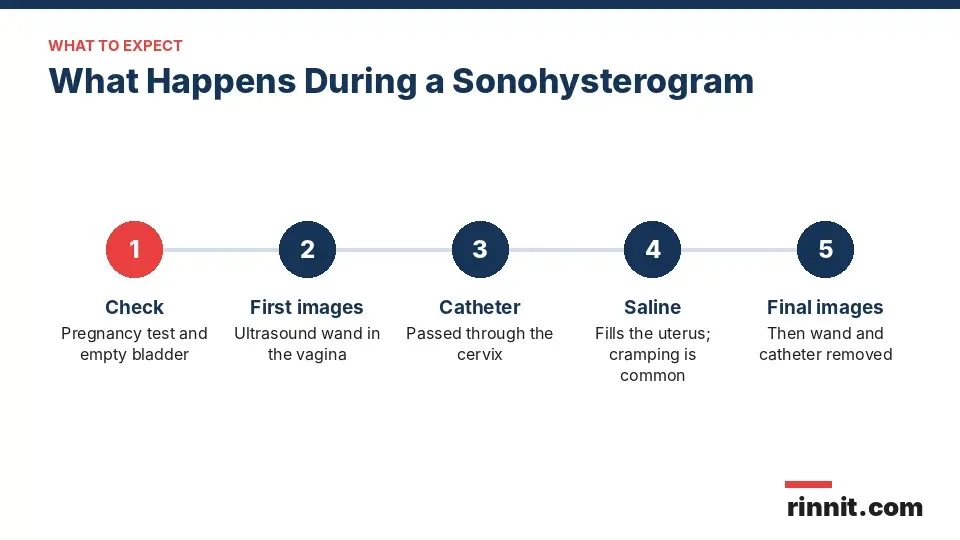
The full visit takes about 30 to 45 minutes, and the saline part lasts about 5 to 10 minutes [1].
Is a sonohysterogram painful?
Mild pain or discomfort is common, especially cramping when the catheter goes in and pressure as the uterus fills [1]. Some people describe mild discomfort and others moderate pain [1]. Cramping usually improves quickly once the fluid drains, and over-the-counter pain relievers and a heating pad can help [1]. Tell your provider if pain becomes intense at any point; you do not have to push through it [1].
Risks of a sonohysterogram
A sonohysterogram is generally safe [1]. Rarely, it can cause a pelvic infection [1]. Contact your provider right away if you have a change in vaginal discharge, a fever or intense pain [1].
After a sonohysterogram
You can usually go home and return to normal activities [1]. A watery discharge a few hours later is the saline draining out [1]. Mild cramping may continue, and pain relievers can help [1]. Light red or brown spotting for a few days can happen from tissue irritation and is usually not a concern [1].
Sonohysterogram results
The test can show [1]:

Depending on the findings, your provider may diagnose a condition, plan surgery to remove a growth, prescribe treatment, monitor a growth or recommend more imaging or tests [1].
Conclusion
A sonohysterogram adds saline to a transvaginal ultrasound to give a clear view of the inside of the uterus. It helps explain irregular bleeding, heavy periods, infertility and repeated miscarriages.
Expect some cramping, take a pain reliever an hour beforehand and call your provider for fever, unusual discharge or severe pain afterward.
Keep learning: Read our guide to the transvaginal ultrasound, our guide to the pelvic ultrasound or our guide to the endometrial biopsy. For more background, see the ACOG guide to sonohysterography.
FAQ
What is a sonohysterogram?
It is a transvaginal ultrasound in which saline is placed in the uterus to give a clearer view of the cavity and lining.
What is another name for a sonohysterogram?
It is also called saline infusion sonography, or SIS.
Why is a sonohysterogram done?
For abnormal or postmenopausal bleeding, heavy or long periods, fibroids, infertility or repeated miscarriages.
When in my cycle should I have a sonohysterogram?
After your period ends and before ovulation, usually cycle days 6 to 11. Some people can have it any time.
How do I prepare for a sonohysterogram?
Eat and take medicines as usual, take a pain reliever an hour before and bring a pad for discharge afterward.
Does a sonohysterogram hurt?
Mild to moderate cramping and pressure are common. Tell your provider if the pain becomes intense.
How long does a sonohysterogram take?
About 30 to 45 minutes in total, with the saline part lasting 5 to 10 minutes.
What is the difference between a sonohysterogram and an HSG?
An HSG uses radiation and dye and can show blocked tubes. A sonohysterogram uses sound waves and saline.
What can a sonohysterogram find?
Polyps, fibroids, a thickened lining, an unusual uterine shape, adenomyosis and signs of endometrial cancer.
What should I expect after a sonohysterogram?
Watery discharge, mild cramps and light spotting for a few days are common. Call for fever, unusual discharge or severe pain.
Related Reading
Uterine Polyps: Symptoms, Causes, and Treatment explains uterine polyps, one of the most common findings on a sonohysterogram.
Adenomyosis: Complete Guide to Symptoms, Causes, Diagnosis & Treatment covers adenomyosis, which this test can show signs of.
What Is Asherman's Syndrome? Symptoms, Causes, and Treatment explains scar tissue inside the uterus that saline sonography can help reveal.
Female Infertility: Causes, Signs, and Treatment Options Explained covers infertility, a common reason to check the uterine cavity with a sonohysterogram.
Heavy Menstrual Bleeding (Menorrhagia): Symptoms, Causes, Diagnosis and Treatment explains heavy periods, which a sonohysterogram is often used to investigate.
Endometrial Cancer: Symptoms, Diagnosis, Treatment and Support covers cancer of the uterine lining, signs of which can show on this test.
References
Cleveland Clinic. Sonohysterogram: Procedure, Fertility, Pain & Side Effects. Cleveland Clinic. Last updated May 23, 2025. https://my.clevelandclinic.org/health/diagnostics/22320-sonohysterogram
de Kroon CD, de Bock GH, Dieben SW, et al. Saline contrast hysterosonography in abnormal uterine bleeding: a systematic review and meta-analysis. BJOG. 2003;110(10):938-947. PMID 14550365. https://doi.org/10.1111/j.1471-0528.2003.02472.x
Seshadri S, El-Toukhy T, Douiri A, et al. Diagnostic accuracy of saline infusion sonography in the evaluation of uterine cavity abnormalities prior to assisted reproductive techniques: a systematic review and meta-analyses. Hum Reprod Update. 2015;21(2):262-274. PMID 25505226. https://doi.org/10.1093/humupd/dmu057
Medical disclaimer
This article is for general education only and does not replace advice from a qualified healthcare professional. Talk to your doctor about your own diagnosis and treatment, and seek emergency care right away for severe or sudden symptoms.



Comments