Pelvic Ultrasound: Types, Preparation, What It Shows and Results

Updated: 2 days ago
Written and medically reviewed by Dr. Baraa Alnahhal, MD · Last reviewed: November 2026 · How we review
Quick Answer: What is a pelvic ultrasound?
A pelvic ultrasound is an imaging test that uses sound waves to make pictures of the organs in your pelvis. It can be done from outside the body (over the belly) or inside (through the vagina or rectum). It helps diagnose problems like cysts, tumors, pain and abnormal bleeding, and it uses no radiation. [1]
What the research says
It reliably rules out endometrial cancer. A meta-analysis of 35 studies and 5,892 women with postmenopausal bleeding found that, at a 5-mm endometrial thickness cutoff, transvaginal ultrasound flagged 96% of women who had endometrial cancer. (Smith-Bindman et al., JAMA, 1998) [2]
A normal scan is reassuring. In the same analysis, a woman with a 10% pre-test chance of endometrial cancer had about a 1% chance after a normal ultrasound. (Smith-Bindman et al., JAMA, 1998) [2]
Expert ultrasound sorts ovarian masses well. Across 47 studies of 19,674 adnexal tumors, expert subjective ultrasound assessment had a sensitivity of 93% and specificity of 89% for ovarian cancer. (Meys et al., European Journal of Cancer, 2016) [3]
Key takeaways
A pelvic ultrasound uses sound waves, not radiation, to image pelvic organs. [1]
It can be done over the belly, through the vagina or through the rectum. [1]
You may need a full bladder for an abdominal scan. [1]
The scan takes 15 minutes to an hour. [1]
It can help diagnose cysts, fibroids, endometriosis, ectopic pregnancy and more. [1]
What organs does a pelvic ultrasound show?
An ultrasound uses sound waves to create detailed pictures called sonograms. A pelvic ultrasound looks at the organs between your abdomen and legs and may also include your lower abdomen. [1]

Bladder, which holds urine
Fallopian tubes, which carry eggs between the ovaries and uterus
Ovaries, which make and store eggs
Prostate, a gland that helps with reproduction in men
Rectum, the lower part of the intestines
Uterus and vagina
Sources: [1]
What are the types of pelvic ultrasound?

Type | How it is done | What it looks at |
Abdominal | The probe moves over the lower belly | Organs seen from outside the belly |
Transvaginal | A probe is placed a few inches into the vagina | Reproductive organs |
Rectal (transrectal) | A probe is placed in the rectum | The rectal lining and the prostate |
Pregnancy | Usually over the belly | The baby's development in the uterus |
Sources: [1]
Why would you need a pelvic ultrasound?
Your provider may recommend one if you have: [1]
Difficulty getting pregnant
Pain during sex or urination
Pelvic or abdominal pain or swelling
Unusual periods or bleeding after menopause
Urinary incontinence (urine leakage)
What can a pelvic ultrasound diagnose?
Group | Conditions |
All people | Bladder cancer, hernias, kidney stones moving toward the bladder |
Females | Ectopic pregnancy, endometriosis, gynecologic tumors, ovarian cysts, ovarian torsion, pelvic inflammatory disease, pelvic organ prolapse, polycystic ovary syndrome (PCOS), uterine fibroids |
Males | Seminal vesicle infections or cysts, prostate cancer, testicular cancer, testicular or scrotal infection, scrotal or penile injury |
Sources: [1]
A pelvic ultrasound can also guide a biopsy needle to the right spot and check the position of an intrauterine device (IUD). [1]
How do you prepare for a pelvic ultrasound?
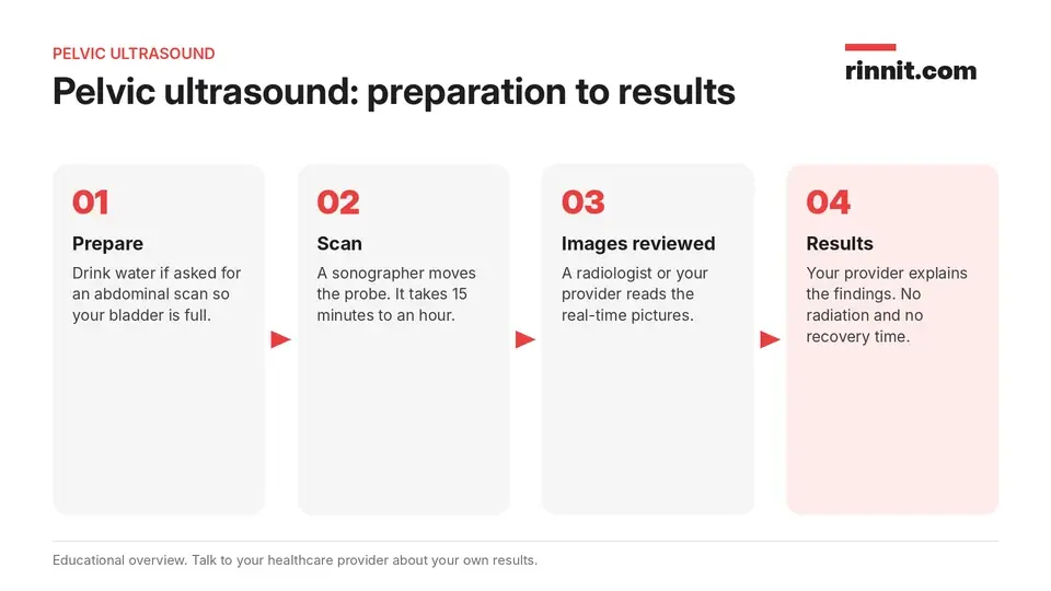
For an abdominal pelvic ultrasound, you may be asked to drink plenty of water first. A full bladder helps the sound waves travel and gives a clearer picture. You usually do not need a full bladder for a transvaginal or rectal ultrasound. [1]
Your provider will give you instructions before the scan, so ask any questions you have. [1]
What happens during a pelvic ultrasound?
A sonographer, a specialist in ultrasound, usually does the scan. They use a small wand called a transducer that sends out sound waves. The echoes bounce back from your tissues and a computer turns them into real-time pictures that can show organ movement and blood flow. [1]
Abdominal: Warm gel goes on your lower belly and the transducer glides over it. You should not feel pain. [1]
Transvaginal: A gel-covered transducer is placed a few inches into the vagina. It may feel a bit uncomfortable at first, like a gynecologic exam. [1]
Rectal: You lie on your side while a lubricated transducer is placed in your rectum to look at its lining and possibly the prostate. [1]
You should not have side effects. Contact your provider right away if you have pain, bleeding, fever or other problems afterward. [1]
How are results shared?
The images usually go to a radiologist, who interprets them and shares a diagnosis with your provider. In obstetrics and gynecology, your provider may read the images directly. Your provider will then contact you to discuss the results. [1]
What are the benefits of a pelvic ultrasound?
Conclusion
A pelvic ultrasound is a safe, quick and reliable way to see the organs in your pelvis. It can find or rule out many conditions, from ovarian cysts to prostate problems, often helping you get treatment sooner or confirming that all is well.
If you have pelvic pain, unusual bleeding or trouble getting pregnant, ask your provider whether a pelvic ultrasound is the right first test.
Keep learning: Read our guide to ovarian cysts or learn about endometriosis. For more background, see RadiologyInfo.org on pelvic ultrasound.
FAQ
What is a pelvic ultrasound?
It is an imaging test that uses sound waves to make pictures of the bladder, reproductive organs and other organs in the pelvis. [1]
What can a pelvic ultrasound detect?
It can help diagnose ovarian cysts, fibroids, endometriosis, ectopic pregnancy, PCOS, hernias, kidney stones and prostate problems. [1]
What are the types of pelvic ultrasound?
The main types are abdominal, transvaginal, rectal (transrectal) and pregnancy ultrasound. [1]
Do I need a full bladder for a pelvic ultrasound?
For an abdominal scan, you may be asked to drink water so your bladder is full; this is usually not needed for transvaginal or rectal scans. [1]
Does a pelvic ultrasound hurt?
An abdominal scan should not hurt; a transvaginal scan may feel a bit uncomfortable at first. [1]
How long does a pelvic ultrasound take?
It takes anywhere from 15 minutes to an hour. [1]
Does a pelvic ultrasound use radiation?
No. It uses sound waves, not radiation. [1]
Who reads the pelvic ultrasound results?
A radiologist usually interprets the images, though your gynecologist may read them directly. [1]
Can men have a pelvic ultrasound?
Yes. It can check the bladder and prostate and help diagnose prostate and testicular problems. [1]
When should I call my doctor after a pelvic ultrasound?
Contact your provider right away if you have pain, bleeding or fever after the scan. [1]
Related Reading
Culdocentesis: Why It's Done, Procedure, Risks and Results explains the needle test that checks fluid behind the uterus.
Ovarian Cysts: Symptoms, Causes, and Treatment Options Explained explains one of the most common findings on pelvic ultrasound.
Endometriosis: Symptoms, Causes, and Treatment covers a condition a pelvic ultrasound can help find.
Polycystic Ovary Syndrome (PCOS): Symptoms, Causes, Diagnosis, and Treatment describes a condition often assessed with pelvic ultrasound.
Ectopic Pregnancy: Symptoms, Causes, and Treatment explains a pregnancy emergency a pelvic ultrasound helps diagnose.
Pelvic Inflammatory Disease: Symptoms, Causes & Treatment — A Clear Guide covers an infection a pelvic ultrasound can help detect.
Ovarian Torsion: Symptoms, Causes, Diagnosis, and Emergency Treatment explains an emergency often diagnosed with pelvic ultrasound.
Endometriosis Ultrasound: What It Can Find, What It Misses, Preparation and Results explains the specialized transvaginal scan that maps endometriomas and deep nodules.
References
Cleveland Clinic. Pelvic Ultrasound: What Is It, Conditions & How It Is Done. Cleveland Clinic. Last updated October 14, 2021. https://my.clevelandclinic.org/health/diagnostics/4997-pelvic-ultrasound
Smith-Bindman R, Kerlikowske K, Feldstein VA, et al. Endovaginal ultrasound to exclude endometrial cancer and other endometrial abnormalities. JAMA. 1998;280(17):1510-1517. PMID 9809732. https://doi.org/10.1001/jama.280.17.1510
Meys EMJ, Kaijser J, Kruitwagen RFPM, et al. Subjective assessment versus ultrasound models to diagnose ovarian cancer: A systematic review and meta-analysis. Eur J Cancer. 2016;58:17-29. PMID 26922169. https://doi.org/10.1016/j.ejca.2016.01.007
Medical disclaimer
This article is for general education only and does not replace advice from a qualified healthcare professional. Talk to your doctor about your own diagnosis and treatment, and seek emergency care right away for severe or sudden symptoms.



Comments