Endometriosis Ultrasound: What It Can Find, What It Misses, Preparation and Results

Updated: 2 days ago
Written and medically reviewed by Dr. Baraa Alnahhal, MD · Last reviewed: November 2026 · How we review
Quick Answer
An endometriosis ultrasound is a detailed transvaginal (and sometimes abdominal) scan that looks for signs of endometriosis, such as endometriomas, deep nodules and adhesions. It takes 30–60 minutes and has no risks. It cannot see superficial lesions smaller than 5 mm, so a normal scan does not rule endometriosis out; laparoscopy remains the definitive test.
What the research says
Ultrasound matches MRI for bowel endometriosis: a meta-analysis of 10 studies and 1,604 women found transvaginal ultrasound detected deep endometriosis of the rectosigmoid bowel with 85% sensitivity and 92% specificity, comparable to MRI at 83% and 95% (Xu Z et al., Frontiers in Medicine, 2025) [2]
It is less sensitive beside the uterus: an updated meta-analysis of 8 studies and 6,728 women found ultrasound detected parametrial involvement with only 63% sensitivity but 98% specificity, so a positive result is reliable while a negative one is not conclusive (Alcázar JL et al., Ultrasound in Obstetrics & Gynecology, 2026) [3]
AI may improve detection of ovarian endometriosis: across 7 studies and 2,737 patients, artificial-intelligence models reading transvaginal ultrasound identified endometriomas with 91% sensitivity and 95% specificity, compared with 80% and 85% for human readers (Mohebbi A et al., Abdominal Radiology, 2026) [4]
Key takeaways
A specialized transvaginal scan that maps endometriomas, deep nodules and adhesions before any surgery.
Can detect deep disease of 5 mm or more in the bowel, bladder and ligaments; misses superficial lesions.
Takes 30–60 minutes; follow instructions about an empty or full bladder.
A positive scan is highly reliable; a negative scan does not exclude endometriosis.
Next steps may include MRI or diagnostic laparoscopy, which confirms the diagnosis with biopsy.
What is an endometriosis ultrasound?
A routine pelvic ultrasound looks at the uterus and ovaries. An endometriosis ultrasound follows a structured protocol that also examines the bowel, bladder, the uterosacral ligaments, the space between the uterus and rectum, and whether the organs move freely against each other (the "sliding sign"). It is performed by a sonographer or gynecologist with specific training in deep endometriosis. Sources: [1]
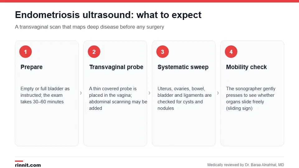
A structured transvaginal scan maps deep disease before surgery.
Why is it done?
Painful periods, pain with sex, bowel or bladder pain around menstruation, or chronic pelvic pain.
Difficulty getting pregnant.
An ovarian cyst that looks like an endometrioma on a routine scan.
Planning surgery: knowing where deep nodules are helps the surgeon choose the team and approach.
Monitoring known disease or the response to hormonal treatment. Sources: [1]
How do I prepare?
Follow the clinic's instruction about your bladder; some protocols want it empty, others partly full, and a bowel preparation is sometimes requested for better views of the rectum.
You can eat normally unless told otherwise.
Scheduling during or just after your period can make small lesions easier to see, but it is not essential.
Tell the team if a transvaginal probe would be painful or unacceptable for you; an abdominal or transrectal approach can be used. Sources: [1]
What happens during the scan?
You undress from the waist down and lie on an exam table with your knees bent.
A thin, covered and lubricated probe is placed in the vagina; an abdominal scan may be added.
The sonographer examines the uterus and ovaries, then sweeps systematically across the bowel, bladder, ligaments and the pouch between the uterus and rectum.
Gentle pressure with the probe and on your abdomen checks whether the ovaries and uterus slide freely over the bowel (sliding sign).
Measurements and images are recorded; the exam takes 30–60 minutes. Sources: [1]
The probe can cause pressure or discomfort, particularly over tender nodules, and you can ask to stop at any time. Sources: [1]
What it can find and what it can miss
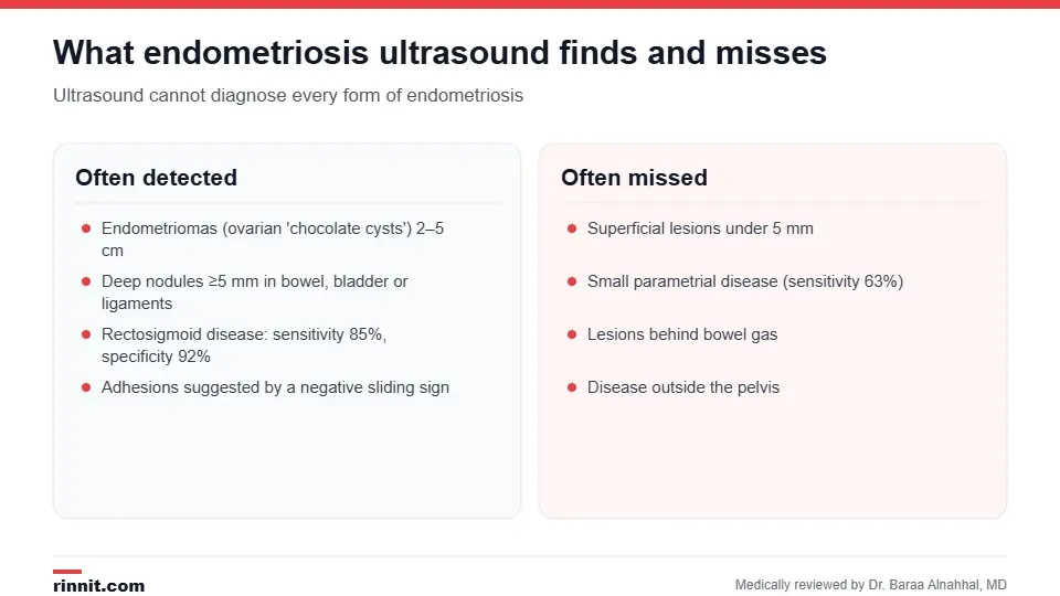
Ultrasound cannot diagnose every form of endometriosis.
Finding | What it looks like | How well ultrasound detects it |
Endometrioma | Ovarian cyst with uniform "ground-glass" contents, usually 2–5 cm | Well detected; AI-assisted reading reaches 91% sensitivity |
Deep endometriosis of the bowel | Thickened, irregular nodule of 5 mm or more in the rectosigmoid wall | Sensitivity 85%, specificity 92% |
Bladder nodule | Solid thickening of the bladder wall | Usually well seen with a partly full bladder |
Uterosacral ligament nodule | Thickened, tender ligament | Moderate |
Parametrial involvement | Tissue beside the cervix | Sensitivity 63%, specificity 98% |
Adhesions | Negative sliding sign | Indirect sign; good specificity |
Superficial peritoneal lesions | Spots under 5 mm on the pelvic lining | Usually not visible |
Sources: [1, 2, 3, 4]
Understanding your results
The report describes each compartment of the pelvis, the size and location of any endometriomas or nodules, and whether the sliding sign is positive or negative. Because the scan is highly specific, a positive finding is reliable and may be enough to start treatment. A normal scan lowers the chance of deep disease but cannot exclude superficial endometriosis, which is only seen at surgery. Results are usually available within a few days. Sources: [1]

A normal scan does not rule endometriosis out.
What happens next?
Your provider may recommend hormonal therapy or pain management based on symptoms and the scan, an MRI to map deep or bowel disease in more detail, or a diagnostic laparoscopy with biopsy, which remains the definitive way to confirm endometriosis. Mapping disease before surgery helps the team plan endometriosis surgery. Sources: [1]
Risks
There are no risks. Ultrasound uses no radiation, and the only side effects are temporary discomfort from the probe and, rarely, light spotting. Sources: [1]
Conclusion
A dedicated endometriosis ultrasound can map endometriomas and deep nodules as well as MRI for the bowel, which makes it a powerful first step before surgery. Its blind spot is superficial disease, so if your scan is normal but symptoms persist, ask about MRI or laparoscopy rather than assuming endometriosis has been excluded.
Keep learning: read our guides to endometriosis and transvaginal ultrasound.
Frequently asked questions
Can an ultrasound diagnose endometriosis?
It can identify endometriomas, deep nodules and adhesions with high specificity, but it cannot see superficial lesions, so it cannot rule endometriosis out. Laparoscopy with biopsy is definitive.
How is it different from a regular pelvic ultrasound?
It follows a structured protocol that also examines the bowel, bladder, ligaments and the sliding of organs, performed by someone trained in deep endometriosis.
Does the scan hurt?
It can be uncomfortable, especially when the probe presses on tender nodules. Tell the sonographer if it is painful; the exam can be paused or adapted.
How long does it take?
Usually 30–60 minutes, longer than a routine pelvic scan because each compartment of the pelvis is checked.
Should my bladder be full or empty?
Follow your clinic's instruction. Some protocols prefer a partly full bladder to see the bladder wall; others prefer it empty for the transvaginal part.
How accurate is ultrasound for bowel endometriosis?
About 85% sensitivity and 92% specificity, comparable to MRI.
What is the sliding sign?
A check of whether the uterus and ovaries slide freely over the bowel. If they do not, adhesions from endometriosis are likely.
What if my scan is normal but I still have symptoms?
Superficial endometriosis is invisible on ultrasound. Your provider may suggest a trial of hormonal treatment, MRI or laparoscopy.
Do I need an MRI as well?
Not always. MRI is added when deep or bowel disease needs more detailed mapping before surgery.
When will I get the results?
Usually within a few days, when your provider reviews the report with you.
Related Reading
Endometriosis: Symptoms, Causes, and Treatment the condition this specialized scan is designed to map.
Transvaginal Ultrasound: Why It's Done, How to Prepare and What to Expect how the probe-based exam works and how to prepare.
Pelvic Ultrasound: Types, Preparation, What It Shows and Results the routine scan that an endometriosis protocol builds on.
Laparoscopy: What It Is, Uses, Procedure and Recovery the keyhole surgery that confirms the diagnosis.
Endometriosis Surgery: Types, Recovery, Risks, and Results how ultrasound mapping guides surgical planning.
References
Cleveland Clinic. Endometriosis Ultrasound: Procedure, Results & Types. Cleveland Clinic. Last updated February 8, 2024. https://my.clevelandclinic.org/health/diagnostics/21814-endometriosis-ultrasound
Xu Z, Li Y, Wang Y, et al. Transvaginal ultrasound and magnetic resonance imaging in detecting rectosigmoid deep infiltrating endometriosis: a comparative meta-analysis. Frontiers in Medicine. 2025;12:1552185. PMID 40166065. https://doi.org/10.3389/fmed.2025.1552185
Alcázar JL, Montoya C, Candau C, et al. Transvaginal ultrasound for detecting parametrial involvement in suspected deep pelvic endometriosis: updated meta-analysis. Ultrasound in Obstetrics & Gynecology. 2026;67(1):7-14. PMID 40848275. https://doi.org/10.1002/uog.70004
Mohebbi A, Zare M, Mohammadi A, et al. Artificial intelligence potential in ovarian endometriosis imaging: a comparative meta-analysis of transvaginal ultrasound-based AI models and human readers. Abdominal Radiology. 2026. PMID 42189210. https://doi.org/10.1007/s00261-026-05570-6
Medical disclaimer
This article is for general education only and does not replace advice from a qualified healthcare professional. Talk to your doctor about your own diagnosis and treatment, and seek emergency care right away for severe or sudden symptoms.



Comments