Renal Scan (Kidney Scan): Types, Preparation, Risks and Results

Written and medically reviewed by Dr. Baraa Alnahhal, MD · Last reviewed: November 2026 · How we review
Quick Answer: What is a renal scan?
A renal scan, or kidney scan, is a nuclear medicine test that shows how well your kidneys work, as well as their shape, size and position. A small amount of radioactive tracer is injected into a vein, and a special camera follows it through your kidneys. [1]
It helps find problems such as blockages, narrowed kidney arteries and poor function after a transplant. It takes 30 minutes to two hours, uses less radiation than an X-ray and carries very low risk. [1]
What the research says
Kidney scans reveal infection damage in children. Pooling 33 studies of children with a first urinary tract infection, 57% had signs of acute kidney infection on a DMSA renal scan and 15% had scarring on a follow-up scan; children with vesicoureteral reflux were 2.6 times as likely to scar. (Shaikh et al., Pediatrics, 2010) [2]
Captopril renography holds its own for renovascular hypertension. A meta-analysis found captopril renography had sensitivity and specificity similar to Doppler and arteriography, was as cost-effective as arteriography and could spare many patients an arteriogram; screening was not cost-effective below a 30% prevalence of renal artery stenosis. (Blaufox et al., Journal of Nuclear Medicine, 1996) [3]
Key takeaways
A renal scan is a nuclear medicine test of kidney function and structure. [1]
A small amount of radioactive tracer is injected into a vein and tracked by a gamma camera. [1]
There are four main types: ACE inhibitor, diuretic, renal cortical and renal perfusion scans. [1]
It uses less radiation than an X-ray, and the risks are very low. [1]
NSAIDs and aspirin can affect results, so share your full medicine list. [1]
What is a renal scan?
A renal scan is a nuclear medicine test that shows how well your kidneys work. It also shows their shape, size and position. A small amount of radioactive material, called a radioisotope or radiotracer, is injected into a vein, usually in your arm or hand. A scanner then finds it in your kidneys and sends images to a computer. [1]
Other names include renal scintigraphy, nuclear medicine renal scan, nuclear medicine kidney scan and renogram. [1]
The images show blood flowing into and out of your kidneys and urine flowing through your kidneys, ureters and bladder. This helps your provider see whether kidney function is reduced or something is blocking urine flow. [1]
Why would you need a renal scan?
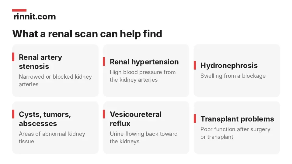
Problems a renal scan can help diagnose.
Providers usually order a renal scan when they suspect your kidneys are not working well, often after blood tests, a urinalysis or X-rays show reduced function. Other reasons include: [1]
An allergy to X-ray dye, which makes a renal scan a safer option
A transplanted kidney
High blood pressure
A kidney blockage that makes urinating difficult or impossible
What can a renal scan diagnose?
Renal hypertension (high blood pressure caused by the kidney arteries)
Hydronephrosis (swollen kidneys from a blockage)
Kidney cysts, tumors or abscesses
Renal artery stenosis (narrowed or blocked kidney arteries)
Poor kidney function after surgery or a transplant
Vesicoureteral reflux (urine flowing back from the bladder toward the kidneys)
Types of renal scans

Each scan type answers a different question.
Type | What it checks | How it's done |
ACE inhibitor renal scintigraphy | Narrowed blood vessels causing high blood pressure | Images before and after an ACE inhibitor medicine |
Diuretic renal scintigraphy | Blockages or problems with urine flow | Images before and after a diuretic (water pill) |
Renal cortical scintigraphy | The outer kidney tissue (renal cortex) | Pictures about two hours after the injection |
Renal perfusion | Blood flow to the kidneys and the width of the kidney arteries | Many pictures over 20 to 30 minutes |
Sources: [1]
How do you prepare for a renal scan?
Make a full list of your medicines, including vitamins and herbal supplements. You may need to stop some a few days before the scan.
Tell your provider if you take NSAIDs such as ibuprofen or aspirin, which can affect results.
Tell your provider about allergies (including drug or latex allergies), claustrophobia or a fear of needles.
Tell your provider if you are breastfeeding, because the tracer may pass into breast milk, or if you could be pregnant, because the scan could harm a fetus.
Drink extra water and empty your bladder before the scan if asked.
Wear comfortable clothes and leave metal items, such as jewelry and eyeglasses, at home.
What happens during a renal scan?
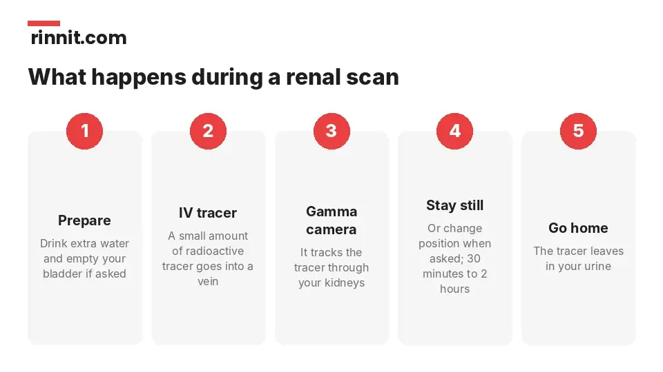
From tracer injection to going home.
A nuclear medicine technologist places an IV, usually in your arm or hand, and injects the tracer.
You lie or sit on an exam table while a gamma camera detects the gamma rays the tracer gives off and creates images of your kidneys.
Depending on the type of scan, you stay still or change position when asked.
The technologist removes the IV, and you can go home.
The scan takes between 30 minutes and two hours, depending on the type. You can return to your usual activities right away. The tracer leaves your body in your urine without discomfort. [1]
Is a renal scan safe?
A renal scan uses less radiation than an X-ray, and the risks are very low. Allergic reactions are rare. The IV site may become discolored, swollen or sore; contact your provider if that discomfort lasts more than a few days. Avoid close contact with children and pregnant women for a few hours afterward. [1]
Contact your provider right away if you notice signs of infection, such as fever, discoloration around the IV site, swelling or headache, or if you think you are having an allergic reaction. [1]
What happens after a renal scan?
A nuclear medicine technologist or radiologist reviews the images and sends a report to your provider, who will discuss the findings with you. If your results are abnormal, your provider may recommend more imaging (ultrasound, MRI or CT), more kidney function tests or a kidney biopsy. [1]
Conclusion
A renal scan shows not just what your kidneys look like but how well they are working, from blood flow to urine drainage. It is especially useful after a transplant, for blockages and when X-ray dye is not an option. [1]
Bring a complete medicine list, mention any chance of pregnancy or breastfeeding, and ask your provider which type of scan you are having and how long to plan for. [1]
Keep learning: Read about renal artery stenosis and vesicoureteral reflux, or see how a kidney biopsy works. For more background, see MedlinePlus on the renal scan.
FAQ
What is a renal scan?
A nuclear medicine test that shows how well your kidneys work and their shape, size and position. [1]
What are other names for a renal scan?
Renal scintigraphy, nuclear medicine renal or kidney scan, and renogram. [1]
Why would I need a renal scan?
To check reduced kidney function, a transplant, high blood pressure or a blockage, or if you are allergic to X-ray dye. [1]
What are the types of renal scans?
ACE inhibitor, diuretic, renal cortical and renal perfusion scans. [1]
How long does a renal scan take?
Between 30 minutes and two hours, depending on the type. [1]
How do I prepare for a renal scan?
Share your medicine list, drink extra water and empty your bladder if asked, and leave metal items at home. [1]
Is a renal scan safe?
Yes. It uses less radiation than an X-ray and the risks are very low. [1]
Can I have a renal scan if I'm pregnant or breastfeeding?
Tell your provider first; the scan could harm a fetus and the tracer may enter breast milk. [1]
What should I avoid after a renal scan?
Avoid close contact with children and pregnant women for a few hours. [1]
What happens if my renal scan is abnormal?
Your provider may order more imaging, kidney function tests or a kidney biopsy. [1]
Related Reading
Narrowed Kidney Arteries: What Renal Artery Stenosis Is, Why It Happens, and How It Is Treated explains the narrowed kidney arteries an ACE inhibitor scan looks for.
What Is Vesicoureteral Reflux? A Plain-Language Guide to the Urine-Backflow Condition in Children covers the urine backflow a renal scan can help diagnose.
Hydronephrosis: What Causes Kidney Swelling, How It Is Found, and How It Is Treated explains kidney swelling from a blockage that a diuretic scan can assess.
Ureteropelvic Junction (UPJ) Obstruction: Causes, Symptoms, and Treatments covers a common blockage where the kidney meets the ureter.
Obstructive Uropathy: Symptoms, Causes, and Recovery explains blocked urine flow that renal scans help measure.
Kidney Transplant Rejection: Understanding the Signs and Prevention covers transplant problems a renal scan can help check.
Renal MAG3 Scan: Kidney Function and Drainage Test Explained explains the nuclear scan that shows how well the kidneys work and drain.
References
Cleveland Clinic. Renal Scan: Purpose, Procedure, Risks & Results. Cleveland Clinic. Last updated April 23, 2025. https://my.clevelandclinic.org/health/diagnostics/21976-renal-scan-kidney-scan
Shaikh N, Ewing AL, Bhatnagar S, et al. Risk of renal scarring in children with a first urinary tract infection: a systematic review. Pediatrics. 2010;126(6):1084-1091. PMID 21059720. https://doi.org/10.1542/peds.2010-0685
Blaufox MD, Middleton ML, Bongiovanni J, et al. Cost efficacy of the diagnosis and therapy of renovascular hypertension. J Nucl Med. 1996;37(1):171-177. PMID 8543990. https://pubmed.ncbi.nlm.nih.gov/8543990/
Medical disclaimer
This article is for general education only and does not replace advice from a qualified healthcare professional. Talk to your doctor about your own diagnosis and treatment, and seek emergency care right away for severe or sudden symptoms.



Comments