Molecular Breast Imaging (MBI): Finding Cancer in Dense Breasts

Updated: 3 days ago
Written and medically reviewed by Dr. Baraa Alnahhal, MD · Last reviewed: November 2026 · How we review
Quick Answer
Molecular breast imaging (MBI) is a nuclear medicine breast test that uses a small amount of radioactive tracer to make fast-growing cells, such as cancer cells, light up on a special camera. It's used along with a mammogram, not instead of one, especially for women with dense breast tissue. The scan takes 20 to 40 minutes.
What the research says
MBI finds more cancers in dense breasts: in 936 women with dense breasts, mammography found 3.2 cancers per 1,000 women, while gamma breast imaging found 9.6 per 1,000, and both together found 10.7 (Rhodes DJ et al., Radiology, 2011) [2]
Low-dose MBI works too: in 1,585 women with dense breasts screened with low-dose MBI, adding it to mammography raised cancer detection from 3.2 to 12.0 per 1,000 and sensitivity from 24% to 91% (Rhodes DJ et al., American Journal of Roentgenology, 2015) [3]
Key takeaways
MBI uses a tracer to highlight fast-growing breast cells.
It's used with mammograms, especially for dense breasts.
The scan takes 20 to 40 minutes with light compression.
Avoid food for 3 hours before; clear fluids are fine.
Highlighted areas may need a biopsy to confirm cancer.
What is molecular breast imaging?
Dense breast tissue looks white on a mammogram, and so do many cancers, so tumors can be hidden. MBI works differently: after a tracer is injected into a vein, cancer cells, which are more active, take up more of it and show as bright spots on a gamma camera, regardless of breast density. It's a type of nuclear medicine imaging. Sources: [1]
Why it's done
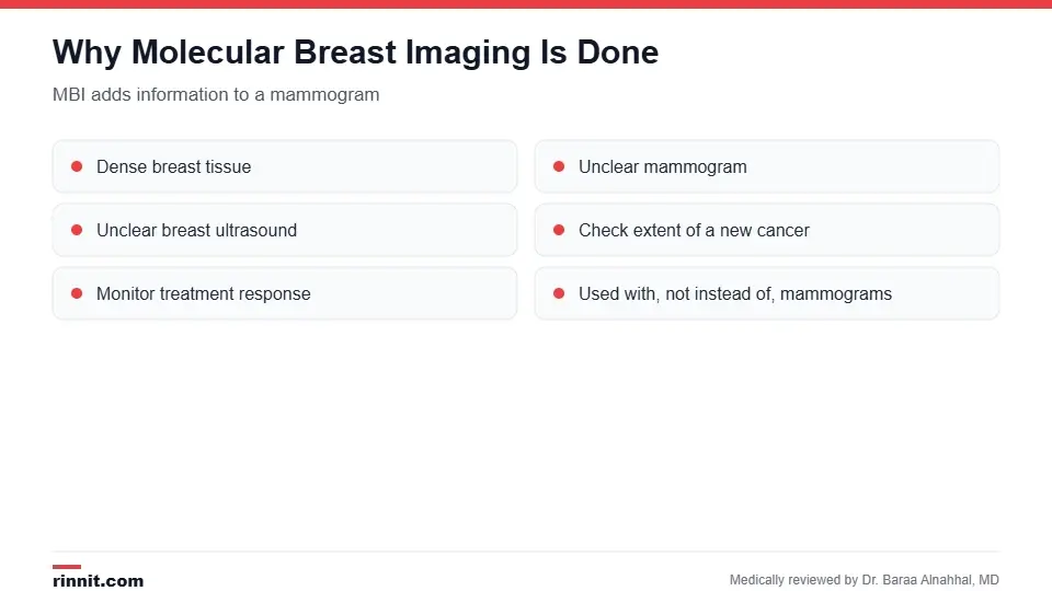
MBI adds information to a mammogram.
Supplemental screening for women with dense breasts.
A mammogram or breast ultrasound that needs more detail.
Checking for other areas of cancer after a diagnosis.
Monitoring how well breast cancer treatment is working. Sources: [1]
MBI vs. other breast imaging
Test | How it works | Role |
Mammogram | Low-dose X-ray | Standard screening |
Layered X-ray images | Better for dense tissue than 2D | |
Magnets and contrast | High-risk screening | |
MBI | Radioactive tracer and gamma camera | Supplemental test, especially for dense breasts |
Sources: [1, 2, 3]
How to prepare
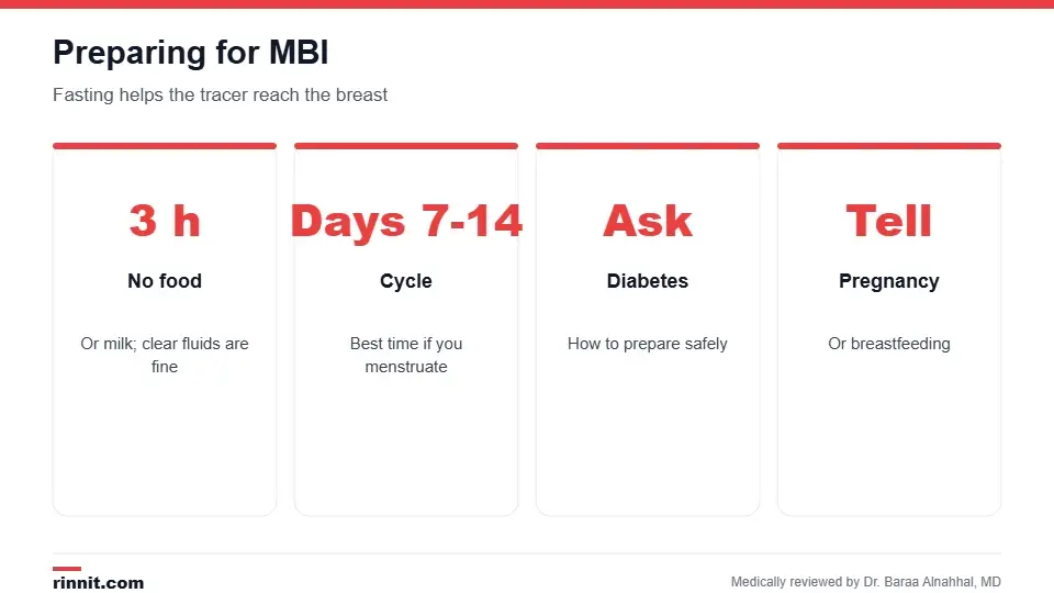
Fasting helps the tracer reach breast tissue.
Don't eat or drink non-clear fluids, such as milk, for at least 3 hours before.
Clear fluids, such as water or sports drinks, are fine.
If you menstruate, schedule the test for days 7 to 14 of your cycle.
If you have diabetes or another condition, ask how to prepare.
Tell your provider if you're pregnant or breastfeeding. Sources: [1]
What happens during the test
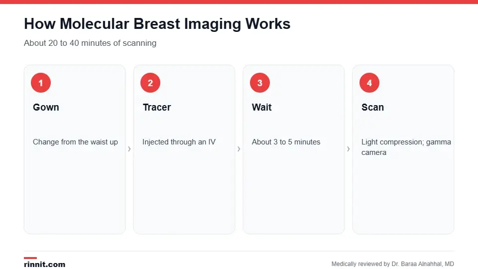
The scan is done sitting in front of the camera.
You change into a gown from the waist up.
You sit in front of a gamma camera, and an IV is placed in your arm.
A small amount of tracer is injected.
After about 3 to 5 minutes, each breast is lightly compressed and imaged, for a total of about 20 to 40 minutes.
The IV is removed and you can get dressed. Sources: [1]
Risks
The radiation dose is low with newer methods, though still a few times higher than a standard mammogram. Allergic reactions to the tracer are rare, affecting about 0.01% to 0.04% of people, and may include hives, itching, rash, nausea or trouble breathing. Sources: [1, 3]
Results
A radiologist reviews the images. No highlighted areas usually means a normal result. A highlighted spot may lead to a breast biopsy to confirm whether it's cancer. In people with suspected breast cancer, MBI detects about 90% of cancers, including 82% of cancers smaller than 10 mm. Results are usually ready within a week. Sources: [1]
Conclusion
Molecular breast imaging can find breast cancers that mammograms miss, especially in dense breasts. Used alongside regular mammograms, it gives women and their care teams a clearer picture.
Keep learning: read about breast cancer screening and the 3D mammogram.
Frequently asked questions
What is molecular breast imaging?
A nuclear medicine test that highlights fast-growing cells in the breast.
Does MBI replace a mammogram?
No. It's used along with mammograms.
Who should consider MBI?
Especially women with dense breast tissue.
How long does MBI take?
About 20 to 40 minutes of scanning.
Do I need to fast?
Yes, avoid food and non-clear fluids for 3 hours.
When should I schedule it?
If you menstruate, on days 7 to 14 of your cycle.
Is MBI painful?
The compression is light, so it's usually more comfortable than a mammogram.
Is the radiation safe?
Yes. The dose is low.
What if a spot lights up?
You may need a biopsy to check it.
When will I get results?
Usually within a week.
Related Reading
Mammogram: Types, How to Prepare, What Happens and Your Results the standard breast screening X-ray.
Breast Cancer Screening: When to Start, How Often and Which Tests Are Used who should be screened and how.
Breast MRI: Why It's Done, What to Expect and How It Compares to a Mammogram another supplemental breast imaging test.
Digital Breast Tomosynthesis (3D Mammogram): Benefits, Prep and Results 3D mammography for dense breasts.
Breast Biopsy: Types, What to Expect, Recovery and Results the test that confirms breast cancer.
References
Cleveland Clinic. Molecular Breast Imaging: What It Is & Procedure Details. Cleveland Clinic. Last updated December 17, 2023. https://my.clevelandclinic.org/health/diagnostics/molecular-breast-imaging
Rhodes DJ, Hruska CB, Phillips SW, et al. Dedicated dual-head gamma imaging for breast cancer screening in women with mammographically dense breasts. Radiology. 2011;258(1):106-118. PMID 21045179. https://doi.org/10.1148/radiol.10100625
Rhodes DJ, Hruska CB, Conners AL, et al. Journal club: molecular breast imaging at reduced radiation dose for supplemental screening in mammographically dense breasts. American Journal of Roentgenology. 2015;204(2):241-251. PMID 25615744. https://doi.org/10.2214/AJR.14.13357
Medical disclaimer
This article is for general education only and does not replace advice from a qualified healthcare professional. Talk to your doctor about your own diagnosis and treatment, and seek emergency care right away for severe or sudden symptoms.



Comments