Intravenous Pyelogram (IVP): What It Is, Why It's Done, Risks and Results

Updated: 2 days ago
Written and medically reviewed by Dr. Baraa Alnahhal, MD · Last reviewed: November 2026 · How we review
Quick Answer: What is an intravenous pyelogram?
An intravenous pyelogram (IVP) is an X-ray test of the urinary system. A provider injects contrast dye into a vein, and X-rays show the dye moving through the kidneys, ureters and bladder. Today IVP is uncommon, because CT scans, MRI and ultrasound usually give more detailed images, but it is still sometimes used for certain kidney conditions. [1]
What the research says
CT has largely replaced IVP for sudden flank pain. In a trial of 224 people with suspected kidney stone pain, 24% of those who had an IVP needed more imaging to reach a diagnosis, compared with 6% after a noncontrast CT. CT took about 30 minutes versus 75 minutes, though it used more radiation (5.0 vs 3.5 mSv). (Thomson et al., Australasian Radiology, 2001) [2]
CT urography is more accurate for finding urinary tract tumors. A review of studies in people with blood in the urine found CT urography had a pooled sensitivity of 96% and specificity of 99% for upper urinary tract tumors, and it outperformed IVP on direct comparison. (Chlapoutakis et al., European Journal of Radiology, 2010) [3]
Key takeaways
An IVP uses contrast dye and X-rays to show how urine travels through your kidneys, ureters and bladder. [1]
It is also called intravenous pyelography, intravenous urography (IVU) or IV urography; IVP and IVU are the same test. [1]
Providers rarely use it now, preferring CT, MRI or ultrasound. [1]
You may get fasting instructions and a laxative, and you pee right before the test. [1]
The test usually takes about an hour. [1]
Mild warmth, flushing or a metallic taste after the dye is common; severe allergic reactions are rare. [1]
What is an intravenous pyelogram?
An IVP is an imaging test that shows how your urinary system handles urine. After the contrast dye is injected into a vein, it travels through your blood to your kidneys. The X-rays pick up the dye, which makes your kidneys, ureters and bladder look white on the images. [1]

Is IVP still used?
Not often. Healthcare providers are now more likely to recommend a CT scan, MRI or ultrasound, which are more comfortable and give more detailed pictures. [1]
What can an IVP help diagnose?
Used in the past for | Still sometimes used for |
Cysts or tumors in the urinary system | Medullary sponge kidney |
Enlarged prostate (benign prostatic hyperplasia) | Renal papillary necrosis |
Kidney stones | |
Urinary problems present from birth | |
Scarring from a urinary tract infection |
Sources: [1]
How do you prepare?
Your provider will review your medical history first. Tell them if you: [1]
Are pregnant or think you might be
Are allergic to skin cleaners such as iodine or isopropyl alcohol
Have had a reaction to contrast dye before
You will get specific instructions about eating and drinking. Your provider may also suggest a laxative, because empty bowels make the images easier to read. [1]
Do you need a full bladder?
No. You should pee right before the test, which makes the dye easier to see in your urinary system. [1]
Who performs the test?
A radiologist (a doctor who specializes in imaging) or a radiologic technologist performs an IVP. [1]
What happens during the test?
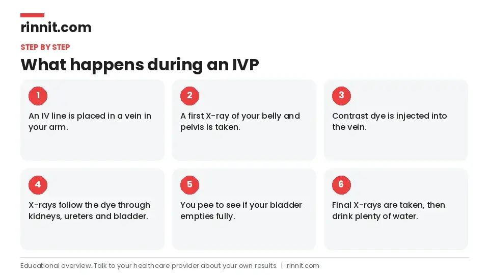
A provider places a small IV line in a vein, usually in your arm. [1]
You lie face-up, and an X-ray of your belly and pelvis is taken before the dye. [1]
The contrast dye is injected into your vein. [1]
Several X-rays are taken as the dye passes through your kidneys, ureters and bladder. [1]
You are asked to pee again to see whether your bladder empties fully. [1]
More X-rays are taken after you pee. [1]
The test usually takes about an hour, and you need to lie still for the images. It can take longer if your kidneys work slowly. [1]
After the test
The IV is removed and a bandage is placed over the site. Drink plenty of fluids, especially water, to help flush the dye out. You can go back to your normal activities right away. [1]
What are the risks?
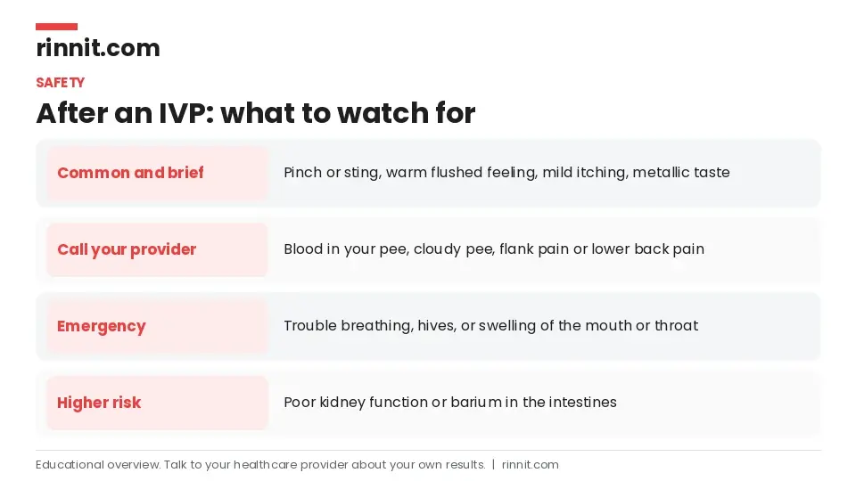
Right after the dye is injected, you may notice short-lived: [1]
A mild pinch or sting
A warm, flushed feeling
Mild itching
A metallic taste in your mouth
Some people feel nauseous or vomit. Severe allergic reactions are rare but possible, with signs such as trouble breathing, low blood pressure, hives, or swelling of the mouth or throat. [1]
Who may not be a good candidate?
What do the results mean?
A radiologist looks for anything unexpected and reports to the provider who ordered the test, who then explains the results and next steps. You can usually expect results within a few days. [1]
An IVP may show: [1]
An unusual shape, size or position of your urinary organs
An enlarged prostate
Blockages
Cysts or tumors
Damage or scarring
If results are abnormal
Your provider may suggest more tests, such as an ultrasound, MRI or CT scan, kidney function tests, or a kidney or bladder biopsy, and may recommend treatment. [1]
When to call your doctor
After an IVP, call your provider if you notice: [1]
Blood in your pee (hematuria)
Cloudy pee
Flank pain
Lower back pain
Go to the nearest emergency room if you have signs of a severe allergic reaction to the contrast dye. [1]
Conclusion
An intravenous pyelogram uses contrast dye and X-rays to show how your urinary system works. Although newer imaging has mostly replaced it, IVP is generally safe, complications are uncommon, and you can return to your usual activities right after the test. [1]
If your provider recommends an IVP, ask why it was chosen over a CT, MRI or ultrasound and what they hope to learn.
Keep learning: Read our guide to the retrograde pyelogram or our guide to kidney stones. For more trusted, doctor-reviewed health guides, visit Rinnit.com.
FAQ
What is an intravenous pyelogram?
It is an X-ray test that uses contrast dye injected into a vein to show your kidneys, ureters and bladder. [1]
Is IVP the same as IVU?
Yes. Intravenous pyelogram and intravenous urogram are two names for the same test. [1]
Why is IVP rarely used today?
CT, MRI and ultrasound are usually more comfortable and give more detailed images. [1]
What can an IVP show?
It can show blockages, stones, cysts, tumors, scarring, an enlarged prostate and unusual organ shape or position. [1]
How do I prepare for an IVP?
Follow eating instructions, take a laxative if advised, pee right before the test, and report any contrast allergy or pregnancy. [1]
How long does an IVP take?
Usually about an hour, sometimes longer if your kidneys work slowly. [1]
Does an IVP hurt?
You may feel a pinch from the IV and a brief warm feeling or metallic taste when the dye goes in. [1]
What are the risks of an IVP?
Mild reactions to the dye are common; severe allergic reactions are rare, and poor kidney function raises the risk of kidney injury. [1]
When will I get my IVP results?
Usually within a few days. [1]
When should I call my doctor after an IVP?
Call if you have blood in your pee, cloudy pee, flank pain or lower back pain, and seek emergency care for a severe allergic reaction. [1]
Related Reading
Kidney Stones: Symptoms, Causes, and Treatment Guide explains kidney stones, one of the main conditions an IVP was used to find.
Understanding Medullary Sponge Kidney: Symptoms, Causes, and Care covers a kidney condition for which providers still sometimes use an IVP.
Renal Papillary Necrosis: Symptoms, Causes, Diagnosis, and Management explains another condition an IVP can still help diagnose.
Benign Prostatic Hyperplasia (BPH): Symptoms, Causes, and Treatment covers an enlarged prostate, which can show up on an IVP.
Urinary Tract Infection (UTI): Symptoms, Causes, Treatment, and Prevention explains UTIs, which can leave scarring visible on this test.
Kidney Cysts: Symptoms, Causes, Diagnosis, and Treatment Options covers cysts in the kidneys that urinary imaging can reveal.
Blood in Urine (Hematuria): Symptoms, Causes, and Treatment explains blood in the urine, a symptom to report to your provider after an IVP.
References
Cleveland Clinic. Intravenous Pyelogram (IVP): Procedure, Risks & Results. Cleveland Clinic. Last updated October 23, 2025. https://my.clevelandclinic.org/health/diagnostics/23578-intravenous-pyelogram
Thomson JM, Glocer J, Abbott C, et al. Computed tomography versus intravenous urography in diagnosis of acute flank pain from urolithiasis: a randomized study comparing imaging costs and radiation dose. Australas Radiol. 2001;45(3):291-297. PMID 11531751. https://doi.org/10.1046/j.1440-1673.2001.00923.x
Chlapoutakis K, Theocharopoulos N, Yarmenitis S, et al. Performance of computed tomographic urography in diagnosis of upper urinary tract urothelial carcinoma, in patients presenting with hematuria: Systematic review and meta-analysis. Eur J Radiol. 2010;73(2):334-338. PMID 19058939. https://doi.org/10.1016/j.ejrad.2008.10.026
Medical disclaimer
This article is for general education only and does not replace advice from a qualified healthcare professional. Talk to your doctor about your own diagnosis and treatment, and seek emergency care right away for severe or sudden symptoms.



Comments