Hip X-Ray: What It Shows, Views Taken and What to Expect

Updated: 2 days ago
Written and medically reviewed by Dr. Baraa Alnahhal, MD · Last reviewed: November 2026 · How we review
Quick Answer
A hip X-ray uses a small dose of radiation to create pictures of the hip joint and nearby pelvic bones. It's often the first test for hip pain, a limp, swelling or an injury, and it can show fractures and arthritis. It takes about 10 minutes. Soft-tissue problems such as a labral tear need MRI or CT instead.
What the research says
X-ray changes and hip pain don't always match: in 946 people from the Framingham study, only 15.6% of hips with frequent pain showed osteoarthritis on X-ray, and only 20.7% of hips with X-ray arthritis were frequently painful; X-ray detected painful hips with just 36.7% sensitivity (Kim C et al., BMJ, 2015) [2]
Some hip fractures are hidden on X-ray: a meta-analysis of older adults with hip pain after a fall and a normal or unclear X-ray found 39% had a fracture needing surgery on advanced imaging, rising to 44% at age 80 or older; MRI was preferred over CT, which missed some fractures (Haj-Mirzaian A et al., Radiology, 2020) [3]
Key takeaways
Shows the hip joint and pelvic bones; often the first test for hip pain or injury.
Finds fractures, arthritis, impingement, infection and hip replacement problems.
At least two views: front (AP) and frog-leg.
Low radiation; tell staff if you might be pregnant.
A normal X-ray doesn't rule out a hidden fracture or soft-tissue injury; MRI may be next.
What is a hip X-ray?
An X-ray passes a small amount of radiation through the body. Dense structures like bone absorb more and appear white; soft tissues appear gray. A hip X-ray focuses on the ball (femoral head) and socket (acetabulum) of the hip joint. A pelvis X-ray shows both hips and the whole pelvis at once. Sources: [1]
Why is it done?

What a hip X-ray can show.
Hip pain, swelling, tenderness or a limp.
A fall or injury, to check for a hip fracture.
Hip arthritis, seen as joint space narrowing and bone spurs.
Hip impingement (FAI), seen as extra bone on the ball or socket.
Avascular necrosis and bone infection.
Hip dysplasia, a shallow hip socket.
Planning and checking a hip replacement. Sources: [1]
How do I prepare?
No special preparation is needed.
Remove jewelry, belts and clothing with metal; you may wear a gown.
Tell the technologist if you are or might be pregnant.
Children receive lower radiation doses. Sources: [1]
What happens during the X-ray?

At least two views are usually taken.
You lie on your back on the X-ray table.
A lead shield may cover nearby parts of your body.
AP view: legs straight and slightly turned in.
Frog-leg view: knees bent and dropped out to the sides, with the soles of your feet together.
You hold still for a few seconds while each image is taken. Sources: [1]
The whole exam usually takes about 10 minutes or a little longer. Sources: [1]
Risks
The radiation dose is low and the benefits usually outweigh the risk. Precautions are taken during pregnancy, and doses are lowered for children. Sources: [1]
Understanding your results
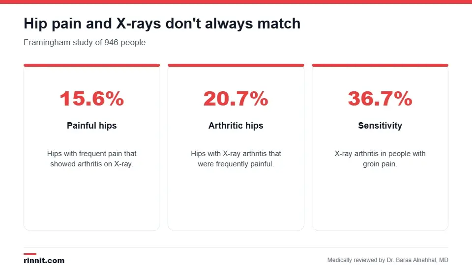
Symptoms and X-ray findings don't always match.
A radiologist reviews the images. In an emergency, results may be available right away; otherwise, usually within 1–2 days. Because X-rays don't show cartilage, the labrum, tendons or every fracture, your provider may order MRI or CT for a suspected labral tear or when a fracture is suspected despite a normal X-ray. Sources: [1, 2, 3]
Conclusion
A hip X-ray is a quick, low-radiation first step for hip pain and injuries, showing fractures, arthritis and bone shape problems. But symptoms and X-ray findings don't always line up, so your provider uses your exam and, if needed, MRI to complete the picture.
Keep learning: read about hip arthritis and hip fractures.
Frequently asked questions
What does a hip X-ray show?
The hip bones and joint, including fractures, arthritis and bone shape problems.
How long does a hip X-ray take?
About 10 minutes or a little longer.
What is a frog-leg view?
An X-ray taken with your knees bent out and the soles of your feet together.
Do I need to prepare?
No. Just remove metal items.
Can a hip X-ray show a labral tear?
No. A labral tear needs MRI or CT.
Can a hip X-ray miss a fracture?
Yes. Some fractures need MRI to be seen.
Is the radiation safe?
Yes, the dose is low. Tell staff if you might be pregnant.
Can a hip X-ray show arthritis?
Yes, as joint space narrowing and bone spurs.
When will I get results?
Right away in emergencies; otherwise within 1–2 days.
Why does my hip hurt if my X-ray is normal?
Pain can come from soft tissues, the spine or early changes X-rays can't show.
Related Reading
Hip Arthritis: Understanding Symptoms, Causes, and Modern Treatments joint wear seen on hip X-rays.
Hip Fracture: Symptoms, Causes, and Treatment breaks diagnosed with hip imaging.
Hip Impingement: Understanding FAI, Symptoms, and Relief extra bone that causes hip pinching.
Hip Replacement Surgery: Procedure, Recovery Time and Risks surgery planned and checked with X-rays.
Avascular Necrosis (Osteonecrosis): Symptoms, Causes, Diagnosis & Treatment — Complete Guide bone death that can affect the hip.
References
Cleveland Clinic. Hip X-Ray: Anatomy & Procedure. Cleveland Clinic. Last updated July 18, 2022. https://my.clevelandclinic.org/health/diagnostics/23508-hip-x-ray
Kim C, Nevitt MC, Niu J, et al. Association of hip pain with radiographic evidence of hip osteoarthritis: diagnostic test study. BMJ. 2015;351:h5983. PMID 26631296. https://doi.org/10.1136/bmj.h5983
Haj-Mirzaian A, Eng J, Khorasani R, et al. Use of advanced imaging for radiographically occult hip fracture in elderly patients: a systematic review and meta-analysis. Radiology. 2020;296(3):521-531. PMID 32633673. https://doi.org/10.1148/radiol.2020192167
Medical disclaimer
This article is for general education only and does not replace advice from a qualified healthcare professional. Talk to your doctor about your own diagnosis and treatment, and seek emergency care right away for severe or sudden symptoms.



Comments