Pelvis X-Ray: What It Shows, What to Expect and When You Need More Imaging

Updated: 2 days ago
Written and medically reviewed by Dr. Baraa Alnahhal, MD · Last reviewed: November 2026 · How we review
Quick Answer
A pelvis X-ray is a painless imaging test that shows the bones of the pelvis. It's used to find the cause of pain, swelling or deformity in the pelvis, hips or upper legs, such as fractures, arthritis and infection. The test takes 10 minutes. Some fractures are hard to see on X-rays, so CT or MRI may follow.
What the research says
A negative X-ray doesn't always rule out a hip fracture: a meta-analysis of 35 studies and 2,992 older adults with suspected hip fracture but no clear fracture on X-ray found that 39% had a hidden fracture needing surgery, rising to 44% in people aged 80 or older; CT detected 79% of these fractures, and the authors favored MRI as the next test (Haj-Mirzaian A et al., Radiology, 2020) [2]
AI may help read hip and pelvic X-rays: in a meta-analysis of 18 studies using AI to diagnose hip fractures on hip or pelvic X-rays, the models had an average sensitivity of 89.3% and specificity of 87.5%, performing comparably to expert radiologists and surgeons (Lex JR et al., JAMA Network Open, 2023) [3]
Key takeaways
Shows the ilium, ischium, pubis, sacrum and coccyx.
Finds fractures, arthritis, infection and alignment problems, and helps plan and check pelvic surgery.
Remove metal; you lie on your back while at least two views are taken (legs straight and legs bent).
Takes about 10 minutes; results usually in one to two days.
Radiation is very low; tell the technologist if you might be pregnant.
What is a pelvis X-ray?
An X-ray machine passes a small amount of radiation through your body to create black-and-white images. Dense bone blocks more radiation and appears white. A pelvis X-ray shows the ring of bones that connects your spine to your legs and supports the organs in your lower belly. A radiologic technologist takes the images, and a radiologist interprets them. Sources: [1]
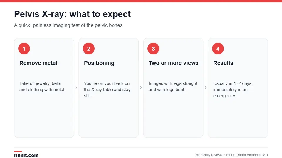
A pelvis X-ray usually takes about 10 minutes.
Why is it done?

A pelvis X-ray shows the bones of the pelvis and hip joints.
Pain, swelling or deformity in the pelvis, hips or upper legs.
Suspected fractures, including hip fractures after a fall.
Hip arthritis and other joint changes.
Bone infection.
Hip dysplasia and alignment problems.
Planning before pelvic surgery and routine checks afterward. Sources: [1]
How do I prepare?
Wear comfortable clothes without metal, or change into a gown.
Remove jewelry, belts and other metal objects.
Tell the technologist if you are or might be pregnant. Sources: [1]
What happens during the X-ray?
You lie on your back on the X-ray table; a lead apron may be placed to shield other areas.
The technologist positions you and steps behind a shield.
At least two views are taken: one with your legs straight (anteroposterior) and one with your legs bent (lateral).
You stay very still and may be asked to hold your breath briefly.
The images are checked, and any blurry ones are retaken. Sources: [1]
The test takes about 10 minutes or more and is painless, though positioning may be uncomfortable if you're injured. Sources: [1]
Understanding your results
In an emergency, results may be available right away. Otherwise, a radiologist usually sends a report to your provider within one to two days. Sources: [1]
Finding | What it may mean |
Break in a bone outline | Pelvic or hip fracture |
Narrowed joint space, bone spurs | Arthritis of the hip or sacroiliac joints |
Areas of bone loss or destruction | Infection or a bone lesion |
Abnormal hip socket shape | Hip dysplasia |
Normal X-ray with ongoing hip pain after a fall | Possible hidden fracture; CT or MRI may be needed |
Pelvis X-rays don't show soft tissues or fluid well; ultrasound or CT is better for those. Sources: [1]
When an X-ray isn't enough
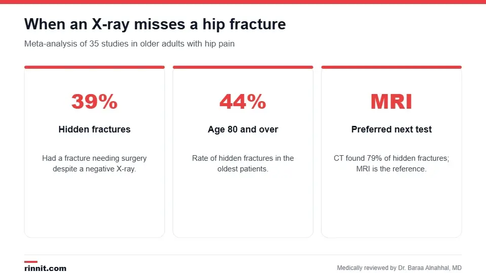
Hidden hip fractures are common in older adults.
Some fractures, especially of the hip in older adults with osteoporosis, can be invisible on the first X-ray. Research shows more than a third of older adults with hip pain after a fall and a negative X-ray have a fracture needing surgery, so providers often order MRI or CT if pain persists or you can't bear weight. AI tools are also being developed to help spot subtle fractures. Sources: [2, 3]
Radiation and safety
The radiation dose is very small and doesn't cause side effects; the very small cancer risk is outweighed by the benefit of a correct diagnosis.
Extra precautions are taken in pregnancy because the pelvis is close to the uterus.
Children are slightly more sensitive to radiation, so lower doses may be used. Sources: [1]
Conclusion
A pelvis X-ray is a fast first look at the bones of your pelvis and hips. It reliably shows most fractures and arthritis, but if you're older, can't bear weight or still have hip pain after a normal X-ray, ask about MRI or CT to rule out a hidden fracture.
Keep learning: read about pelvis anatomy and tailbone pain.
Frequently asked questions
What does a pelvis X-ray show?
The hip bones (ilium, ischium and pubis), sacrum, tailbone and hip joints.
How long does a pelvis X-ray take?
About 10 minutes or more.
Does a pelvis X-ray hurt?
No, though positioning can be uncomfortable if you're injured.
How many views are taken?
At least two: legs straight and legs bent.
Can a pelvis X-ray miss a fracture?
Yes. Hidden hip fractures are common in older adults, so MRI or CT may be needed.
Is a pelvis X-ray safe in pregnancy?
Extra precautions are needed; tell the technologist if you might be pregnant.
When will I get my results?
Usually within one to two days, or immediately in an emergency.
Can it show arthritis?
Yes. Narrowed joint spaces and bone spurs suggest arthritis.
Does a pelvis X-ray show organs?
Not clearly. Ultrasound, CT or MRI is used for organs and fluid.
Can it tell a male pelvis from a female pelvis?
Yes. The female pelvis is broader with a pubic arch over 90 degrees; the male pelvis is narrower with an arch under 90 degrees.
Related Reading
Pelvis: Anatomy, Bones, Function, Shapes and Common Conditions the bones a pelvis X-ray shows.
Hip Fracture: Symptoms, Causes, and Treatment a fracture that may need more than an X-ray to find.
Hip Arthritis: Understanding Symptoms, Causes, and Modern Treatments joint changes seen on pelvis X-rays.
Hip Dysplasia: Symptoms, Causes, Diagnosis & Treatment — Complete Guide a hip socket problem diagnosed with X-rays.
Coccydynia: Tailbone Pain Causes, Symptoms, Treatment, and Relief tailbone pain that may lead to imaging.
References
Cleveland Clinic. Pelvis X-ray: Purpose, Procedure & Risks. Cleveland Clinic. Last updated July 18, 2022. https://my.clevelandclinic.org/health/diagnostics/23519-pelvis-x-ray
Haj-Mirzaian A, Eng J, Khorasani R, et al. Use of advanced imaging for radiographically occult hip fracture in elderly patients: a systematic review and meta-analysis. Radiology. 2020;296(3):521-531. PMID 32633673. https://doi.org/10.1148/radiol.2020192167
Lex JR, Di Michele J, Koucheki R, et al. Artificial intelligence for hip fracture detection and outcome prediction: a systematic review and meta-analysis. JAMA Network Open. 2023;6(3):e233391. PMID 36930153. https://doi.org/10.1001/jamanetworkopen.2023.3391
Medical disclaimer
This article is for general education only and does not replace advice from a qualified healthcare professional. Talk to your doctor about your own diagnosis and treatment, and seek emergency care right away for severe or sudden symptoms.



Comments