Ureteral Obstruction: Symptoms, Causes, and Treatment

Updated: 3 days ago
Medically reviewed by Dr. Baraa Alnahhal, MD. Last reviewed October 17, 2026. This content is for educational purposes and is not medical advice.
Quick Answer: What is a ureteral obstruction?
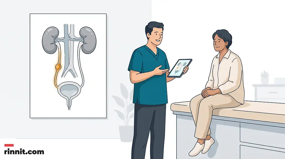
A ureteral obstruction is a blockage in one or both ureters that stops urine from draining from the kidneys into the bladder [3]. Warning signs include flank or back pain, changes in urine output [3], blood in the urine and recurrent infections. It is often found on ultrasound [3]. Treatment involves urgent drainage with a stent or tube, then surgery for the blockage [3] [5].
What the research says
For cancer-related blockages, stents have practical advantages over nephrostomy tubes. A meta-analysis of 10 comparative studies found that stents were associated with shorter procedure time (mean difference −10.26 minutes), shorter hospital stay (mean difference −1.30 days; 95% CI −1.69 to −0.92), and fewer accidental tube dislodgments (OR 0.25; 95% CI 0.13 to 0.48), with no difference in overall complications or survival (Gauhar et al., International Braz J Urol, 2022) [6].
When a blocked kidney is infected, the type of drainage may matter. In a comparison of 65 patients needing emergency drainage for acute upper urinary tract obstruction with urosepsis (35 nephrostomy, 30 retrograde stent), postoperative C-reactive protein and the time to a normal temperature were significantly lower with nephrostomy, especially in patients with severe inflammation and fever (Xu et al., Journal of Infection and Chemotherapy, 2021) [7].
Minimally invasive surgery appears as effective as open surgery for blockages in infants. In a meta-analysis of 11 studies of pyeloplasty for ureteropelvic junction obstruction in infants, minimally invasive surgery took longer but shortened hospital stay, with no significant difference in complications (OR 0.84; 95% CI 0.52 to 1.35) or success rate (OR 1.35; 95% CI 0.59 to 3.07) (Wang et al., PeerJ, 2023) [8].
How urine is supposed to flow — and what goes wrong
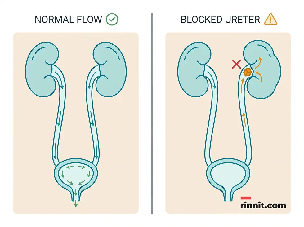
The urinary system has four main parts: the kidneys, the ureters, the bladder, and the urethra. The kidneys sit toward the back of the upper abdomen and filter waste and fluid from the blood to make urine. That urine then travels down narrow tubes called the ureters, which empty into the bladder. The bladder stores urine until you urinate, and the urethra carries it out of the body.
A ureteral obstruction is a blockage in one or both of those tubes. The kidneys keep making urine even when it cannot escape, so pressure builds up behind the blockage. Over time, that pressure can cause the kidney to swell (a condition doctors call hydronephrosis) [3] and stop working properly.
Here is the part that matters most: ureteral obstruction can be cured, and severe complications are rare because it is treatable. But the timeline is not forgiving. Left untreated, symptoms can move quickly from mild — pain, fever, and infection — to severe — loss of kidney function, sepsis, and death. That is why warning signs deserve prompt medical attention, not a wait-and-see approach.
Ureteral obstruction symptoms: what to watch for
A ureteral obstruction can be completely silent. Whether symptoms appear depends on where the blockage sits, whether it is partial or complete, how fast it developed, and whether one kidney or both are affected. Some people discover a blockage only on a routine scan.
When symptoms do appear, they usually follow this pattern:
Symptom | What it typically looks like |
Pain | Often in the back, side, or flank [3]; may be sharp or come in waves |
Changes in urine output | You may produce noticeably more or less urine than usual [3] |
Difficulty urinating | Urine flow feels weak, interrupted, or hard to start |
Blood in the urine | Pink, red, or brown urine, which may appear without pain |
Urinary tract infections | Repeated infections that keep returning after treatment |
High blood pressure | New or unexplained hypertension linked to kidney pressure [3] |
When to seek urgent care. Pain is the most common alarm, but the combination of symptoms matters more than any single one. Get medical attention right away if you experience:
Fever or chills alongside a suspected blockage deserve particular urgency. A blocked kidney that becomes infected can progress to sepsis [3], a body-wide infection that is life-threatening.
Ureteral obstruction causes: why the tube gets blocked
Different types of ureteral obstruction have different causes, and some are present at birth (congenital) [3]. The blockages fall into two broad groups: things inside the tube (intrinsic) and things pressing on it from outside (extrinsic).
Causes present at birth
A second (duplicated) ureter. This common congenital condition means two ureters form on the same kidney. The second ureter may be fully or only partially developed. If either tube does not work properly, urine can back up into the kidney and cause damage.
A blockage where the ureter meets the kidney or bladder. [3] These two junctions are the most common pinch points. A blockage at the kidney end (the ureteropelvic junction) can cause the kidney to swell and eventually stop working. It may be present at birth, develop during normal childhood growth, result from injury or scarring, or, in rare cases, develop from a tumor. A blockage at the bladder end (the ureterovesical junction) causes urine to back up into both the tube and the kidney.
Ureterocele. When a ureter is too narrow to let urine flow through completely, a tiny bulge can form in the tube, usually at the end closest to the bladder. That bulge can itself block flow and force urine back into the kidney.
Causes that develop later
Retroperitoneal fibrosis. This rare disorder causes fibrous tissue to grow in the space behind the abdominal organs. The fibers can develop as a result of cancer tumors or from taking certain medicines used to treat migraines. The tissue encircles and blocks the ureters [3], causing urine to back up.
Other intrinsic and extrinsic causes. A wider set of factors can narrow or pinch a ureter at any point along its length:
Cause | How it blocks the ureter |
Kidney stones | A stone lodges in the narrow tube and physically stops urine flow [3] |
Cancerous and noncancerous tumors | |
Blood clots | Clots obstruct flow within the tube [3] |
Enlarged lymph nodes | Swollen nodes press on the ureter from outside |
Internal tissue growth | Conditions such as endometriosis in females can compress the tube |
Long-term ureter wall swelling | Chronic inflammation from diseases such as tuberculosis [3] or the parasite infection schistosomiasis |
Who is at higher risk. Certain conditions present at birth raise the chance of a blockage. Kidney stones and bladder stones also increase risk, as do blood clots, tumors, certain tissue growths, and enlarged lymph nodes.
The stakes. An untreated obstruction can lead to urinary tract infections and kidney damage — and the damage can be irreversible [3]. This is the single most important reason to act on the warning signs rather than assuming the pain will pass.
How a blocked ureter is diagnosed
Some ureteral obstructions are found before birth. [3] Routine prenatal ultrasounds can show details of the developing fetus, including the kidneys, ureters, and bladder. Doctors often order another ultrasound after birth to recheck the kidneys.
For older children and adults, if a provider suspects an obstructed ureter, several tests and scans may be used to confirm the diagnosis:
Diagnostic test | What it does |
Blood and urine tests | Check for signs of infection and measure creatinine [3], which rises when the kidneys are not working properly |
Ultrasound | A retroperitoneal ultrasound images the kidneys and ureters [3] without radiation |
Bladder catheterization with X-rays | A small tube is passed through the urethra, dye is injected into the bladder, and X-rays capture the kidneys, ureters, bladder, and urethra before and during urination [3] |
Renal nuclear scan | A tracer with a small amount of radioactive material is injected into the arm; a camera detects it to evaluate how well the urinary system drains [3] |
Cystoscopy | A small camera tube is inserted into the urethra or through a small incision so the doctor can see inside the urethra and bladder directly [3] |
CT scan | X-ray views from many angles are combined into cross-sectional images of the kidneys, ureter, and bladder [3] |
MRI | An abdominal MRI uses a magnetic field and radio waves to image the organs and tissues of the urinary system in detail [3] |
Ureteral obstruction treatment: draining first, fixing second
The goal of treatment is straightforward: remove the blockage if possible, or bypass it [3]. Either approach can help repair damage to the kidneys. If an infection is present, antibiotics are used to clear it.
Drainage procedures: the urgent first step
An obstruction causing severe pain may need an immediate procedure to remove urine from the body and temporarily relieve the pressure [3]. A urinary tract specialist (urologist) may recommend one of three options:
A ureteral stent — a hollow tube inserted inside the ureter to keep it open so urine can flow again [3] [5].
Percutaneous nephrostomy — a tube inserted through the back to drain the kidney directly [3] [5].
A catheter — a tube passed through the urethra that connects the bladder to an external drainage bag. This can matter especially when bladder problems also contribute to poor kidney drainage.
Your urologist will tell you which procedure or combination works best for your situation. Drainage procedures may provide temporary or permanent relief [3], depending on the underlying cause.
Surgical procedures: fixing the blockage
Once urine is draining, the blockage itself can be addressed. Several surgical procedures correct ureteral obstructions, and the choice depends on your specific situation:
Surgical approach | How it works |
Endoscopic surgery | A lighted scope passes through the urethra into the bladder and urinary tract; the surgeon widens the blocked or damaged part of the ureter and places a stent to keep it open; the same session can diagnose and treat |
Open surgery | The surgeon makes an incision in the abdomen to remove the blockage and repair the ureter |
Laparoscopic surgery | One or more small incisions allow a lighted tube with a camera and instruments to remove or repair the blockage |
Robot-assisted laparoscopic surgery | The surgeon guides a robotic system to perform the laparoscopic procedure |
The main differences among these approaches are recovery time and the number and size of the incisions. Your urologist determines which procedure and approach best fits your condition.

Preparing for your appointment
Depending on how frequent and severe your symptoms are, you may start with your primary care provider or be referred directly to a urologist. Preparation makes that visit far more productive.
Before the appointment, ask whether you need to do anything in advance, such as fasting before a specific test. Then build four lists: your symptoms (including anything that seems unrelated), key personal information (major stresses, recent life changes, and family medical history), all medications, vitamins, and supplements with their doses, and the questions you want to ask. If possible, bring a family member or friend to help remember what is discussed.
Questions worth asking include: What is likely causing my symptoms? What other causes are possible? What tests do I need? Is my condition likely temporary or chronic? What is the best course of action, and what alternatives exist? How do my other health conditions affect the plan? Are there restrictions I should follow? Should I see a specialist?
What your provider will ask: When did your symptoms begin? Have they been continuous or occasional? How severe are they? What improves them, and what makes them worse?
In the meantime, avoid anything that seems to worsen your signs and symptoms, and treat any fever or severe pain as a reason to seek care promptly rather than wait.
Conclusion
A ureteral obstruction is a plumbing problem with serious consequences if it is ignored. The blockage itself is often curable, but the kidney behind it does not tolerate pressure well: repeated infections and irreversible kidney damage are the real risks, and sepsis is the worst-case endpoint. The good news is that modern diagnosis — from routine prenatal ultrasound to CT, MRI, and nuclear scanning — catches most blockages early, and modern treatment drains the kidney quickly and fixes the underlying cause through stents, nephrostomy tubes, or minimally invasive surgery.
If you have flank or back pain that will not settle, changes in your urine output, blood in your urine, or repeated urinary tract infections, do not wait it out. See a healthcare provider promptly, and treat fever with severe pain as an urgent situation.
Frequently asked questions
Can a blocked ureter go away on its own? Sometimes a small kidney stone passes through the ureter without intervention, and some blockages found before birth resolve after delivery. But a persistent obstruction will not resolve by itself and can damage the kidney, so a blockage should always be evaluated by a healthcare provider rather than left to chance.
What does ureteral obstruction pain feel like? The pain often occurs in the back, side, or flank and can be severe enough that you cannot sit still or find a comfortable position. When it comes with nausea and vomiting, or with fever and chills, it signals an urgent need for medical attention.
Can a ureteral obstruction cause kidney damage? Yes. When urine cannot drain, pressure builds on the kidney and can cause it to swell. Repeated infections and prolonged pressure can lead to kidney damage that may be irreversible, which is why prompt drainage is a treatment priority.
Is a ureteral stent permanent? Not usually. A stent is often a temporary measure that keeps the ureter open while the underlying cause is treated or while the kidney recovers. Depending on the condition, drainage procedures can provide temporary or permanent relief, and your urologist will decide which is appropriate for you.
Can ureteral obstruction be detected before birth? Yes. Routine prenatal ultrasounds can show the developing kidneys, ureters, and bladder and often detect an obstruction before delivery. A repeat ultrasound is usually performed after birth to recheck the kidneys.
Trusted External Resources
Related Reading
Ureteral Stent: Why It's Used, Placement, Side Effects and Removal explains ureteral stents and what to expect.
Nephrostomy Tube: Purpose, Placement and Care explains nephrostomy tubes and how to care for them.
Learn more in our guide to Extracorporeal Shock Wave Lithotripsy (ESWL): Procedure, Recovery, Risks and Results.
CT Urogram: What It Is, Why It's Done, How to Prepare and What to Expect explains the contrast CT scan of the urinary system used to look for this condition.
References
UpToDate. Clinical manifestations and diagnosis of urinary tract obstruction (UTO) and hydronephrosis. UpToDate. Accessed October 30, 2026. https://www.uptodate.com/contents/clinical-manifestations-and-diagnosis-of-urinary-tract-obstruction-uto-and-hydronephrosis
Yu ASL, et al., eds. Urinary tract obstruction. In: Brenner & Rector's The Kidney. 11th ed. Elsevier; 2020. Accessed October 9, 2021.
Preminger GM. Obstructive Uropathy. Merck Manual Professional Edition. Last updated August 2026. https://www.merckmanuals.com/professional/genitourinary-disorders/obstructive-uropathy/obstructive-uropathy
Mayo Clinic Staff. Ureteral obstruction - Symptoms and causes. Mayo Clinic. December 23, 2025. https://www.mayoclinic.org/diseases-conditions/ureteral-obstruction/symptoms-causes/syc-20354676
Tabib C, Nethala D, Kozel Z, et al. Management and treatment options when facing malignant ureteral obstruction. Int J Urol. 2020;27(7):591-598. PMID 32253785. https://doi.org/10.1111/iju.14235
Gauhar V, Pirola GM, Scarcella S, et al. Nephrostomy tube versus double J ureteral stent in patients with malignant ureteric obstruction. A systematic review and meta-analysis of comparative studies. Int Braz J Urol. 2022;48(6):903-914. PMID 36037256. https://doi.org/10.1590/S1677-5538.IBJU.2022.0225
Xu ZH, Yang YH, Zhou S, et al. Percutaneous nephrostomy versus retrograde ureteral stent for acute upper urinary tract obstruction with urosepsis. J Infect Chemother. 2021;27(2):323-328. PMID 33309627. https://doi.org/10.1016/j.jiac.2020.11.022
Wang M, Xi Y, Huang N, et al. Minimally invasive pyeloplasty versus open pyeloplasty for ureteropelvic junction obstruction in infants: a systematic review and meta-analysis. PeerJ. 2023;11:e16468. PMID 38025670. https://doi.org/10.7717/peerj.16468



Comments