Understanding the Uvea: The Eye’s Essential Middle Layer

Updated: 3 days ago
Medically reviewed by Dr. Baraa Alnahhal, MD · Last reviewed: October 2026
Editorial note: This article is provided for educational purposes only. It is not medical advice. Always consult a qualified healthcare professional about your individual health situation.
Quick Answer: What is the uvea?
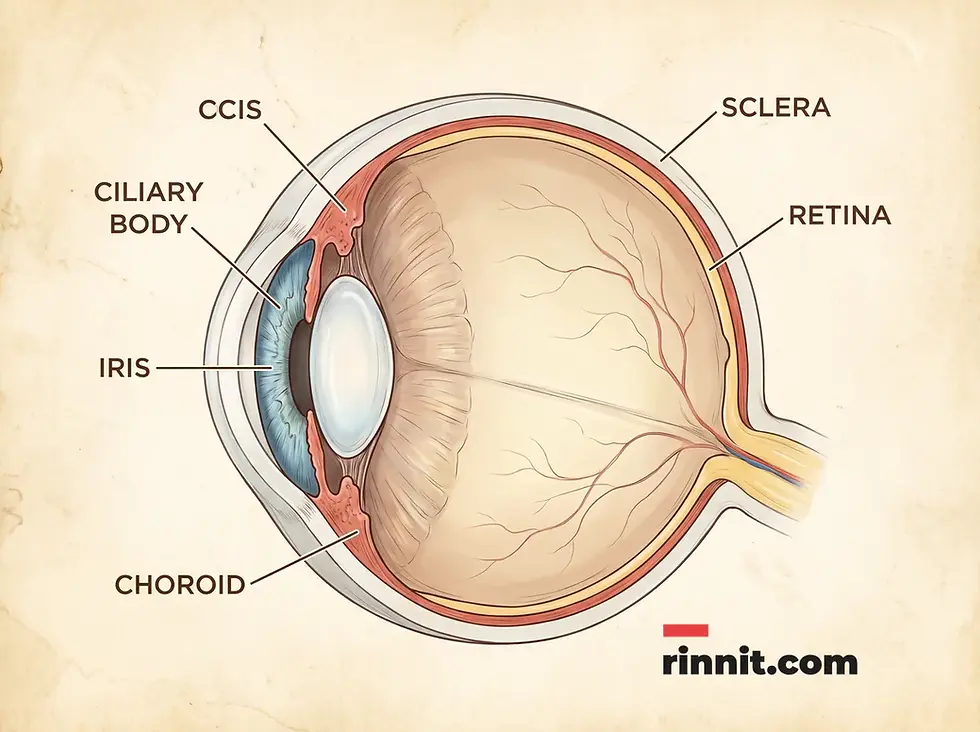
The uvea is the middle layer of the eye's wall, between the sclera and the retina, also called the vascular tunic because parts are rich in blood vessels. [1] It has three parts: the iris, which controls light and gives eye color, the ciliary body, which focuses the lens, and the choroid, which supplies the retina. [1] Uveitis can cause vision loss. [4]
What the research says
Most uveitis has no single identified cause. Pooling 12 U.S. studies of 11,146 patients, undifferentiated uveitis accounted for 42.18% of cases (95% CI 32.68–52.29%); among defined causes, HLA-B27-associated uveitis (5.65%) and sarcoidosis (4.98%) were the most common non-infectious causes, and toxoplasmosis (4.07%) was the leading infectious cause (Liba et al., Seminars in Ophthalmology, 2026) [2].
The choroid is the retina’s main supply line, and its blood flow tracks with heart and vessel health. A review found that the choroid supplies the bulk of the retina’s oxygen and nutrients, that coronary artery disease and carotid artery stenosis are linked to decreased choroidal thickness and blood flow, and that carotid endarterectomy may reverse these changes (Yeung et al., Survey of Ophthalmology, 2020) [3].
Uveitis can cause lasting complications, especially when it starts in childhood. In a French cohort of 147 children (268 affected eyes) with chronic or recurrent non-infectious uveitis, 161 complications occurred, including permanent visual loss in 31 patients, although many cases became inactive after biologic treatments were introduced (Morelle et al., RMD Open, 2019) [4].
When to See a Doctor
Outside regular eye exams, see an eye care specialist for eye symptoms that last longer than a few days, disrupt your usual activities, or involve any kind of vision loss. [1] Don’t ignore what feels like an eye infection for more than a few days, because infections can spread and turn into bigger problems. [1]
Sudden vision loss — such as blurring, clouding, or other sudden changes — is always a medical emergency. Call emergency services immediately. [1]
Anatomy and Function of the Uvea
The uvea is the middle of three layers in the eye’s outer wall. Its name comes from Latin and Ancient Greek words meaning “grape,” because from the outside most of the uvea resembles a grape’s skin. [1] Another name, the “vascular tunic,” reflects that it wraps around most of the eye like a shirt and that parts of it are packed with blood vessels. [1]
The uvea isn’t one tissue but three, each with its own job:
Uveal part | Main function | Key feature |
Iris | Controls how much light enters the eye | Gives the eye its color [1] |
Ciliary body | Shapes the lens for focusing and makes aqueous humor [1] | A ring of tissue just behind the iris |
Choroid | Supplies blood to the eye, especially the retina | Largest part of the uvea; contains melanin [1] |
The Iris and Ciliary Body: Light and Focus
The iris is a ring of muscle tissue just behind the cornea that automatically widens or narrows to control how much light enters your eyes. [1] Just behind it, the ciliary body’s muscles control the shape of the lens, helping you focus on objects at different distances. [1] The ciliary body also makes aqueous humor, a fluid that fills part of the eye and helps it hold its globe-like shape. [1]

The Choroid: The Eye’s Plumbing
The choroid is a thin sheet of tissue packed with blood vessels, wrapping around the central and rear sections of the eyeball, just outside the retina and just inside the sclera. [1] Cleveland Clinic compares the eye to a house: the choroid is the plumbing, bringing blood to different places, especially the retina. [1]
The choroid also contains a lot of melanin, the pigment that gives skin and eyes their color. Researchers suspect it helps absorb some of the light entering the eye, reducing how much light reflects off the retina. [1]

Common Conditions and Symptoms
Because the uvea has several parts with different jobs, it is vulnerable to several types of conditions. These include albinism, choroideremia, coloboma, diabetes-related retinopathy, eye cancers (especially intraocular melanoma), eye ischemia (lack of blood flow, such as from an eye stroke), injuries, iris malformations such as corectopia or polycoria, macular degeneration (especially the wet form), presbyopia, sarcoidosis, synechiae, and uveitis. [1]
Uveitis, inflammation of the uvea, can lead to complications such as glaucoma and retinal problems, so it is usually managed with specialist input. Symptoms depend on which part of the uvea is affected:
Symptom category | Common signs |
Visual changes | Blurred vision, double vision, sudden or gradual vision loss [1] |
Pupil changes | Dilated or pinpoint pupils, or a pupil that changes from round to irregular [1] |
Discomfort | Eye pain, eye strain, headache, light sensitivity (photophobia) [1] |
Surface signs | Red eye or irritation, watery eyes, swollen conjunctiva [1] |
Accumulations | White liquid at the bottom of the front of the eye or in a ring around the iris (hypopyon) [1] |
Protecting Your Uvea
Care step | What to do | Why it matters |
Regular eye exams | Get an exam every one to two years, or more often if you have a condition that needs it [1] | Detects many eye conditions before symptoms appear |
Eye protection | Wear goggles or safety glasses made for impact hazards, even if you wear glasses [1] | Prevents injuries or limits how severe they are |
Avoid nicotine | Avoid smoking, vaping, and smokeless tobacco [1] | Protects the delicate blood vessels of the uvea |
Don’t ignore infections | See a provider if an apparent eye infection lasts more than a few days [1] | Prevents spread and bigger problems |
Conclusion
The uvea is a quiet but indispensable part of the eye. By controlling light, focusing the lens, and supplying blood to the retina, its three parts keep vision working. It is vulnerable to a range of conditions, but regular exams and good protective habits can catch problems early.
Understanding the uvea’s role makes it easier to recognize symptoms such as pain, light sensitivity, redness, and pupil changes, and to act quickly when they appear.
Your next step: Schedule a comprehensive eye exam if you haven’t had one in the past one to two years. If you notice eye pain, light sensitivity, redness that lasts more than a few days, or any vision loss, see an eye care specialist. Treat sudden vision loss as an emergency.
Related reading: Uveitis | Panuveitis | The Iris | Synechiae of the Eye | The Anterior Chamber | The Macula
Frequently Asked Questions
1. What is the uvea?
The uvea is the middle of the three layers that make up the eye’s outer wall, and it contains the iris, ciliary body, and choroid.
2. Why is it called the vascular tunic?
“Vascular” means circulation-related, and a tunic is a type of shirt. The uvea wraps around most, but not all, of the eye, and parts of it are packed with blood vessels.
3. What does the iris do?
The iris is a ring of muscle tissue at the front of the eye, just behind the cornea. It widens or narrows automatically to control how much light enters the eye, and it gives the eye its color.
4. What is the function of the ciliary body?
Muscles in the ciliary body control the shape of the lens so you can focus on objects around you, and the ciliary body makes the aqueous humor fluid that helps the eye hold its globe-like shape.
5. What role does the choroid play?
The choroid is a thin sheet of tissue packed with blood vessels that supplies blood to parts of the eye, especially the retina. It is the largest part of the uvea.
6. What does the word “uvea” mean?
The word “uvea” comes from Latin and Ancient Greek words meaning “grape,” because from the outside most of the uvea looks like the skin of a grape.
7. What is uveitis?
Uveitis is inflammation of the uvea. It can cause eye pain and vision loss, and it can happen for unknown reasons or because of many known conditions.
8. Can albinism affect the uvea?
Yes. Albinism is among the conditions that can affect the uvea.
9. What is photophobia?
Sensitivity to light. It is one of the symptoms that can occur with uveal conditions.
10. What can an irregularly shaped pupil mean?
A change in pupil shape from a circle to an irregular shape can be a symptom of a uveal condition, so it deserves a prompt evaluation by an eye care specialist.
11. How often should I get an eye exam?
Everyone should get an eye exam every one to two years, or more often if you have a condition that needs closer monitoring.
12. Does smoking affect the uvea?
Smoking, vaping, and smokeless tobacco can all affect the circulatory system, including the delicate blood vessels in the uvea.
13. What is hypopyon?
White liquid that collects at the bottom of the front of the eye, or in a ring around the iris. It is a symptom of some uveal conditions.
14. Is sudden vision loss an emergency?
Yes. Sudden vision loss, such as blurring, clouding, or other changes, is always a medical emergency, and you should call emergency services immediately.
15. How can I protect my uvea from injury?
Wear goggles or safety glasses made to handle flying-object impacts or other hazards, even if you already wear glasses.
16. What causes uveitis?
Often no cause is found. In pooled U.S. studies, about 42% of cases were undifferentiated, and among defined causes HLA-B27-associated uveitis, sarcoidosis, and toxoplasmosis were most common.
References
Cleveland Clinic. Uvea. Cleveland Clinic. August 6, 2024. https://my.clevelandclinic.org/health/body/uvea
Liba T, Olami A, Cabir Y, et al. The etiological spectrum of uveitis in the United States: a systematic review and meta-analysis. Semin Ophthalmol. 2026 Aug 26:1-11. PMID 42655880. https://doi.org/10.1080/08820538.2026.2721691
Yeung SC, You Y, Howe KL, Yan P. Choroidal thickness in patients with cardiovascular disease: a review. Surv Ophthalmol. 2020;65(4):473-486. PMID 31923478. https://doi.org/10.1016/j.survophthal.2019.12.007
Morelle G, Gueudry J, Uettwiller F, et al. Chronic and recurrent non-infectious paediatric-onset uveitis: a French cohort. RMD Open. 2019;5(2):e000933. PMID 31452929. https://doi.org/10.1136/rmdopen-2019-000933
Medical disclaimer
This article is for education only and is not a substitute for professional medical advice, diagnosis, or treatment. Sudden vision loss or sudden changes in vision are medical emergencies. Always consult a qualified healthcare professional about your individual health situation.



Comments