Synechiae of the Eye: Causes, Symptoms, and Vision Protection

Updated: 2 days ago
Medically reviewed by Dr. Baraa Alnahhal, MD · Last reviewed: October 2026
TL;DR: Synechiae of the eye are abnormal adhesions where the iris sticks to the lens or the cornea due to scar tissue or inflammation. [1] This condition can distort the pupil, cause significant eye pain, and lead to serious complications like glaucoma if the flow of fluid within the eye is blocked. Early diagnosis through specialized eye exams and treatment with medicated drops or surgery are essential to prevent permanent vision loss.
Quick Answer: What are synechiae of the eye?
Synechiae of the eye are abnormal adhesions in which the iris sticks to the lens behind it or the cornea in front of it, typically after inflammation or injury. [1] They can raise eye pressure and lead to glaucoma. [1] Symptoms include eye pain, redness and changes in pupil shape. Treatment includes corticosteroids, pupil-dilating drops or surgery in severe cases.
What the research says
Glaucoma raises the stakes in inflamed eyes. An international expert group notes that uveitic glaucoma carries nearly three times the risk of severe visual loss compared with uveitis without glaucoma. Its consensus recommendations call for a stricter pressure target of 16 mmHg and early referral to a glaucoma specialist (Pantcheva et al., Ocul Immunol Inflamm, 2025) [2].
Steroids calm inflammation but can raise eye pressure. In a Cochrane review of two trials (401 people with chronic non-infectious uveitis), a steroid implant probably prevented uveitis from returning (risk ratio 0.29, 95% CI 0.14–0.59) but raised the need for cataract surgery (risk ratio 2.98, 95% CI 2.33–3.79) and pressure-lowering surgery (risk ratio 7.48, 95% CI 3.94–14.19) (Brady et al., Cochrane Database Syst Rev, 2016) [3].
Newer pressure-lowering drops are being studied. A meta-analysis of 11 studies (383 participants) found that Rho-kinase inhibitor drops lowered eye pressure in uveitic glaucoma by about 9.3 mmHg at 3 months (95% CI −17.0 to −1.6) and 7.9 mmHg at 12 months (95% CI −10.3 to −5.6). Five of the 11 studies were at high risk of bias (Aguilar-Barrera et al., Graefes Arch Clin Exp Ophthalmol, 2026) [4].
What are Synechiae of the Eye?
Ocular synechiae occur when tissues within the eye adhere to one another in locations where they should remain separate. Specifically, the iris—the colored membrane that controls the size of the pupil—becomes stuck to the clear structures located either directly in front of or behind it. These adhesions are typically the result of scar tissue formation following an injury, surgery, or prolonged inflammation. [1]
When these tissues stick together, they can interfere with the normal movement of the iris and the flow of aqueous humor, the clear fluid that nourishes the eye and maintains its internal pressure. If left unmanaged, the resulting pressure buildup can cause irreversible damage to the optic nerve. [1]

What are the types of synechiae of the eye?
Ocular synechiae are classified based on which structure the iris has adhered to. Both types can impact vision and eye health, though one is more common than the other.
Type | Description | Frequency and Impact |
|---|---|---|
Posterior Synechiae | The iris adheres to the lens, the transparent structure located behind it. [1] | The most common form; can lead to a fixed or distorted pupil. |
Anterior Synechiae | The iris adheres to the cornea, the clear outer covering of the front of the eye. [1] | Can significantly interfere with the eye's drainage system. |
What are the symptoms and warning signs of synechiae of the eye?
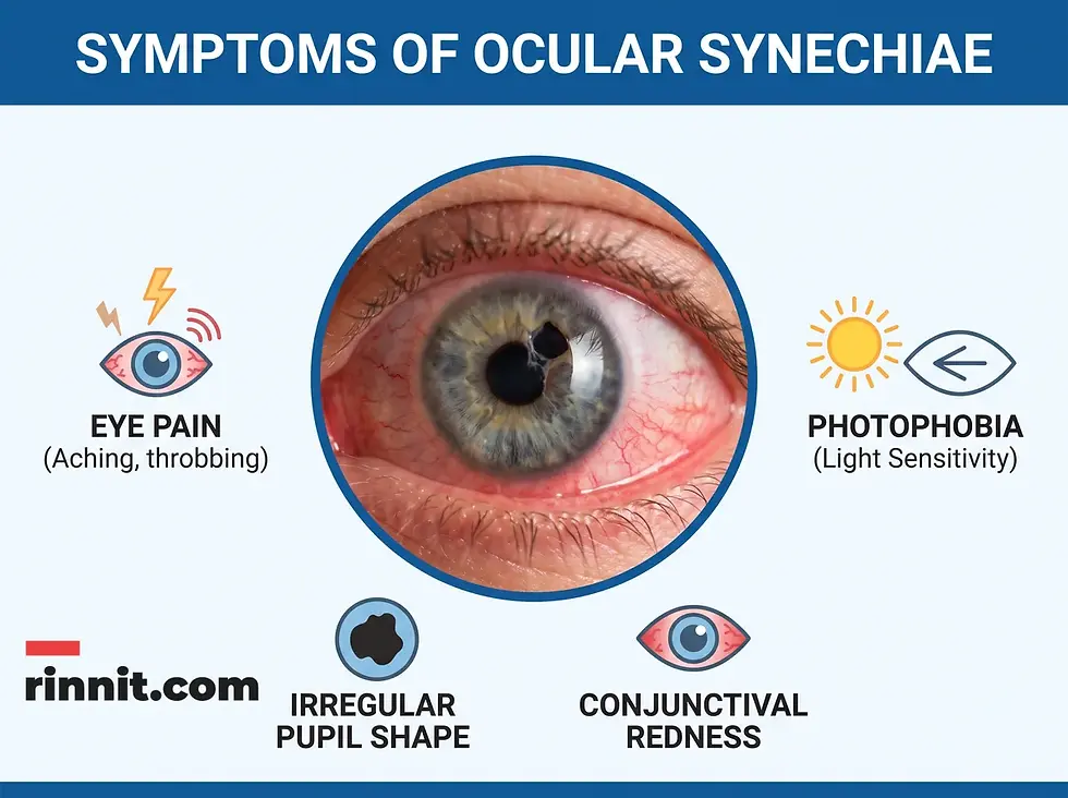
The symptoms of synechiae often overlap with other inflammatory eye conditions, making a professional assessment critical. Because the iris is directly involved, visible changes to the center of the eye are common.
Pupil Changes: The pupil may appear abnormally small (miosis), abnormally large (mydriasis), or irregular in shape rather than perfectly round. [1]
Physical Discomfort: Patients often experience aching, irritation, or sharp pain within the eye.
Visual Sensitivity: Increased sensitivity to light (photophobia) and watery eyes are frequent complaints. [1]
Visible Redness: The white part of the eye may appear bloodshot or inflamed.
Vision Quality: Blurring or a noticeable loss of vision can occur as the condition progresses.
What causes synechiae of the eye, and who is at risk?
Most cases of synechiae are acquired rather than congenital. They are almost always a secondary response to another issue affecting the eye's delicate internal environment.
Inflammatory Diseases: Conditions like uveitis (inflammation of the middle layer of the eye) or iritis (inflammation of the iris) are primary drivers of adhesion formation. [1]
Trauma and Injury: Physical damage to the eye can trigger the formation of scar tissue as the eye attempts to heal.
Surgical Complications: Adhesions can sometimes develop as a side effect of eye surgeries, such as those performed to treat cataracts.
Congenital Factors: In rare instances, some individuals are born with ocular synechiae. [1]
How are synechiae of the eye linked to glaucoma?
The most serious complication associated with synechiae is the development of glaucoma. The eye relies on the constant flow of aqueous humor to provide nutrition and maintain a healthy level of intraocular pressure. Synechiae act as a physical barrier, blocking the channels through which this fluid moves. [1]
When the fluid cannot drain properly, pressure inside the eye rises rapidly. This elevated pressure puts direct stress on the optic nerve, which transmits visual information to the brain. If the optic nerve is damaged by this pressure, it can lead to permanent vision loss or total blindness. [1]
How are synechiae of the eye diagnosed?
Diagnosing synechiae requires a comprehensive eye examination by a specialist. Several tests are used to visualize the adhesions and measure their impact on eye function.
Slit-Lamp Exam: A specialized microscope is used to provide a high-magnification, three-dimensional view of the iris, lens, and cornea.
Tonometry: This test measures the internal pressure of the eye to check for signs of glaucoma. [1]
Visual Acuity and Field Tests: Reading charts and peripheral vision tests help determine if any vision loss has already occurred.
Optical Coherence Tomography (OCT): This imaging technology creates detailed cross-sectional images of the tissues at the back of the eye.
How are synechiae of the eye managed and treated?
Treatment for synechiae is focused on two goals: breaking the existing adhesions and preventing new ones by controlling inflammation. The specific approach depends on the severity of the case and the presence of complications like glaucoma.
Treatment Category | Common Methods |
|---|---|
Pupil Dilation | Mydriatic eye drops are used to enlarge the pupil and relax iris muscles to help separate the tissues. [1] |
Inflammation Control | Corticosteroids in the form of drops, ointments, or pills help reduce swelling and scar tissue. [1] |
Pressure Management | Glaucoma medications, such as beta-blockers, are prescribed to lower intraocular pressure. [1] |
Surgical Intervention | Lasers or small incisions may be used to drain fluid or separate severe adhesions. |

What is the long-term outlook for synechiae of the eye?
The prognosis for individuals with eye synechiae is generally positive if the condition is diagnosed and treated in its early stages. [1] Most cases can be managed effectively with medication, preventing the onset of glaucoma. However, because the risk of recurrence is higher in those with chronic inflammatory conditions, ongoing monitoring by an eye care specialist is often necessary to ensure long-term vision protection.
Conclusion
Synechiae of the eye are scar-like adhesions that form when the iris sticks to the lens or cornea, most often after inflammation, injury, or surgery. The danger lies in blocked fluid drainage and rising eye pressure, which can damage the optic nerve. Early specialist care, inflammation control, and regular pressure checks give the best chance of protecting your sight.
What are the next steps for synechiae of the eye?
If you notice changes in the shape of your pupil, persistent eye pain, or increased sensitivity to light, do not wait. Schedule a comprehensive eye exam today to ensure any underlying adhesions are identified and treated before they impact your vision. Get emergency care right away for sudden severe eye pain with blurred vision, halos around lights, headache, nausea or vomiting, which can signal a sudden rise in eye pressure.
Related reading on Rinnit: Uveitis: Symptoms, Causes, and Treatment — What Eye Inflammation Means for Your Vision · Angle-Closure Glaucoma: An Eye Emergency Caused by Blocked Fluid Drainage · Cataracts: Symptoms, Causes, Diagnosis and Treatment — What You Need to Know · Goniotomy: A Microinvasive Approach to Glaucoma Treatment
Frequently Asked Questions
Can synechiae heal on their own?
No, adhesions caused by scar tissue typically require medical intervention to be broken or managed.
Is surgery always necessary for synechiae?
No, many cases are successfully treated with specialized eye drops and anti-inflammatory medications.
What is the difference between anterior and posterior synechiae?
Posterior involves the iris sticking to the lens, while anterior involves the iris sticking to the cornea.
Can synechiae cause blindness?
If left untreated, they can lead to glaucoma, which can cause permanent vision loss or blindness.
Are synechiae painful?
Yes, they are often accompanied by eye aching, irritation, and sensitivity to light.
Can cataracts cause synechiae?
Yes, cataracts and the surgeries used to treat them are known risk factors for developing adhesions.
What are mydriatic drops?
Mydriatic drops are eye drops that dilate the pupil and relax the muscles of the iris.
How does inflammation lead to synechiae?
Chronic inflammation can cause the production of fibrin and other sticky substances that form scar tissue.
Can children get eye synechiae?
Yes, they can be present at birth (congenital) or develop due to injury or pediatric inflammatory conditions.
Will my vision return to normal after treatment?
If caught early, vision usually remains stable. If damage has already occurred, treatment focuses on preventing further loss.
How often should I have my eye pressure checked?
Your eye care specialist will determine a schedule based on your specific risk factors for glaucoma.
What is uveitis?
Uveitis is inflammation of the middle layer of the eye and is a major cause of ocular synechiae.
Can eye injuries from years ago cause synechiae now?
While they usually develop shortly after an injury, chronic inflammation following trauma can lead to later adhesions.
Are there lifestyle changes that prevent synechiae?
Managing underlying inflammatory conditions is the most effective way to prevent them.
Can synechiae return after surgery?
Yes, especially if the underlying cause of inflammation is not fully controlled.
References
Cleveland Clinic. Synechiae (Eye). Cleveland Clinic. March 29, 2023. https://my.clevelandclinic.org/health/diseases/24874-synechiae-eye
Pantcheva MB, Gangaputra S, Sieck E, et al. Uveitic Glaucoma Interest Group recommendations for uveitis-related ocular hypertension and glaucoma management. Ocul Immunol Inflamm. 2025;33(9):2153-2167. PMID 40971800. https://doi.org/10.1080/09273948.2025.2542286
Brady CJ, Villanti AC, Law HA, et al. Corticosteroid implants for chronic non-infectious uveitis. Cochrane Database Syst Rev. 2016;2(2):CD010469. PMID 26866343. https://doi.org/10.1002/14651858.CD010469.pub2
Aguilar-Barrera B, Mejía-Salgado G, Cardona-López J, et al. Efficacy, effectiveness, and safety of rho-kinase inhibitors in uveitic glaucoma and ocular hypertension secondary to uveitis: a systematic review and meta-analysis. Graefes Arch Clin Exp Ophthalmol. 2026;264(6):1527-1540. PMID 41632210. https://doi.org/10.1007/s00417-025-07111-1
Medical Disclaimer
This article is for informational purposes only and does not constitute medical advice. Always seek the advice of your physician or other qualified health provider with any questions you may have regarding a medical condition.



Comments