Understanding the Iris: Anatomy, Function, and Care

Updated: 3 days ago
Medically reviewed by Dr. Baraa Alnahhal, MD | Last reviewed: October 7, 2026
Quick Answer: What is the iris?
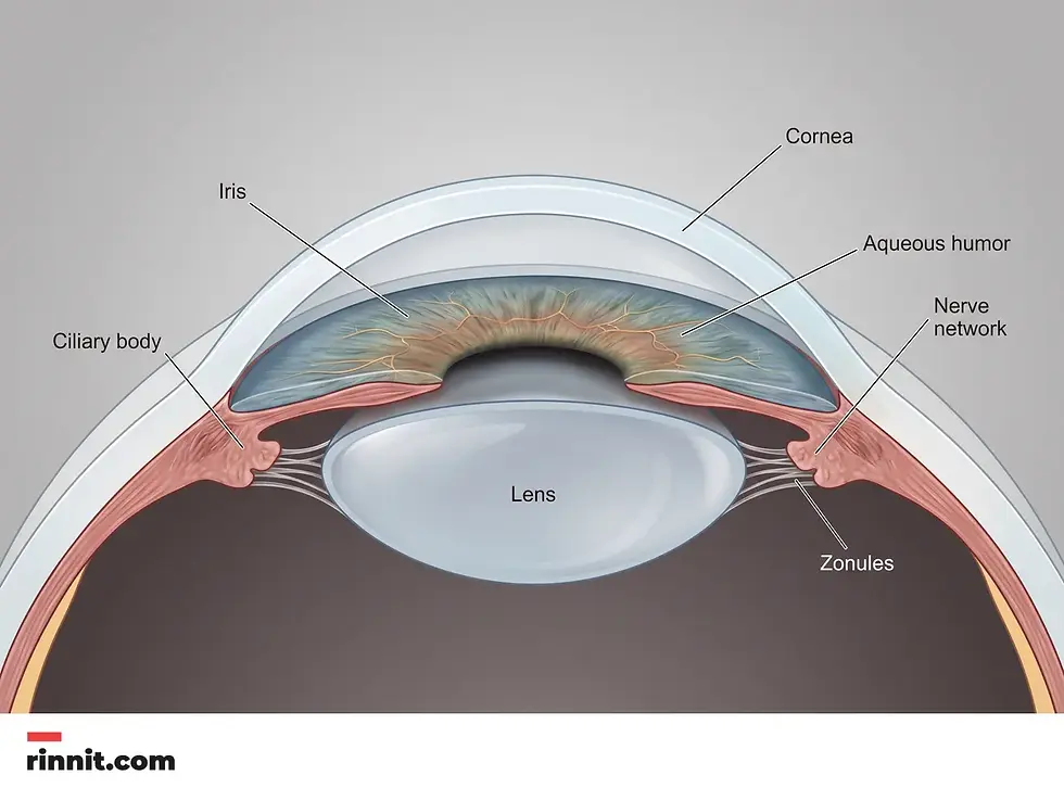
The iris is the flat, circular, colored part of the eye located behind the cornea and in front of the lens [1]. It regulates how much light enters the eye by automatically adjusting pupil size [1], using specialized muscles to widen the pupil in low light and narrow it in bright light. This supports clear vision and protects internal eye structures.
What the research says
Glaucoma, an eye condition that can involve the iris’s drainage angle, affects about 1 in 28 people aged 40–80 worldwide, and cases are rising. A systematic review and meta-analysis of 50 population-based studies estimated a global glaucoma prevalence of 3.54% (95% credible interval 2.09–5.82) in people aged 40–80, equal to 64.3 million people in 2013, rising to 76.0 million in 2020 and a projected 111.8 million in 2040 (Tham et al., Ophthalmology, 2014) [2].
More sunlight exposure is linked to a higher risk of age-related cataract, which is why UV protection is recommended. A meta-analysis of 31 epidemiological studies found that people with the highest sunlight exposure had a pooled odds ratio of 1.15 (95% CI 1.09–1.21) for age-related cataract, with a small but significant link for cortical cataract (pooled OR 1.03, 95% CI 1.02–1.03) and none for nuclear or posterior subcapsular cataract (Li et al., BioMed Research International, 2021) [3].
Uveitis, which can inflame the iris, is uncommon but estimates vary widely. A systematic review and meta-analysis of 49 studies found a pooled uveitis incidence of 50.45 per 100,000, with prevalence estimates ranging from 9 to 730 cases per 100,000 depending on the population, definitions and region (García-Aparicio et al., Ophthalmic Epidemiology, 2021) [4].
The Role and Function of the Iris
The iris serves as the eye’s natural light regulator, working continuously to balance visual clarity with ocular protection. This process occurs automatically through the autonomic nervous system [1], meaning individuals do not have to consciously think about adjusting their vision when moving between different lighting environments.
When a person enters a dimly lit room, the muscles within the iris adjust to widen the pupil, a process known as dilation [1]. This allows more light to reach the retina, enhancing the ability to see in the dark. Conversely, in bright sunlight, the iris muscles narrow the pupil [1], preventing an overwhelming amount of light from entering and potentially damaging sensitive internal tissues.
Anatomical Structure and Composition
Located between the clear cornea and the crystalline lens, the iris is a complex tissue composed primarily of muscles and nerves [1]. It is often compared to a fingerprint [1] because the intricate patterns and colors are unique to every individual, determined by a combination of genetics and melanin levels passed down from biological parents [1].
The iris is one of several layers that form the eyeball [1], sitting relatively close to the surface. Its color is not merely aesthetic; it is the result of how much pigment, or melanin, the body produces [1]. Higher concentrations of melanin typically result in darker eye colors, such as brown, while lower levels result in lighter shades like blue or green.
Table 1: Structural Characteristics of the Iris
Feature | Description |
Shape | Flat, round, and circular [1] |
Location | Under the cornea and on top of the lens [1] |
Primary Tissues | Muscles and nerves that work automatically, without conscious control [1] |
Unique Identifier | Unique patterns similar to a fingerprint |
Pigmentation | Determined by melanin levels and genetic factors [1] |
Common Conditions and Disorders
While the iris is a resilient structure, it can be affected by various medical conditions, congenital disorders, and physical injuries. Some issues are present from birth, while others may develop due to age, systemic health problems, or trauma to the eye.
Conditions such as aniridia involve the total or partial absence of the iris, which significantly impacts light regulation. Inflammatory issues like uveitis can cause pain and vision changes, while systemic syndromes like Waardenburg syndrome may affect pigmentation. It is important to note that any significant change in the appearance or function of the iris should be evaluated by a medical professional.
Table 2: Medical Conditions Affecting the Iris
Condition | Impact on the Iris and Eye |
Aniridia | The partial or complete absence of the iris tissue |
Uveitis | Inflammation of the middle layer of the eye, including the iris |
Glaucoma | Increased eye pressure that can damage ocular structures |
Albinism | Reduced pigmentation affecting eye color and light sensitivity |
Horner’s Syndrome | A nerve-related condition affecting pupil dilation |
Cataracts | Clouding of the lens located directly behind the iris |

Diagnostics and Professional Care
Medical professionals use a variety of tools to assess the health of the iris during comprehensive eye examinations. A common method is the slit lamp exam [1], which uses a high-intensity light source and a microscope to provide a detailed view of the iris and other anterior structures of the eye.
If a specific disorder is suspected, more advanced imaging may be required [1]. These tests help identify structural abnormalities or internal damage that might not be visible during a standard physical checkup.
Diagnostic Testing Methods
Slit Lamp Exam: Provides a magnified, three-dimensional view of the iris.
Optical Coherence Tomography (OCT): Uses light waves to take cross-section pictures of the tissue.
Ultrasound: Employs sound waves to visualize the internal structures of the eye.
Imaging Scans: MRI and CT scans may be used for complex neurological or structural assessments.
Proactive Maintenance and Safety
Maintaining the health of the iris involves both protective habits and regular professional monitoring. One of the most effective ways to protect the iris is by wearing high-quality sunglasses. Protective eyewear should be labeled as providing 100% UV protection or UV400 [1], which shields the eyes from harmful ultraviolet radiation.
Additionally, modern technology often utilizes iris scanners for security purposes. Current medical evidence suggests that the infrared light used in these devices is safe and does not cause damage to the eye or its internal components [1].

Conclusion
The iris is a remarkable anatomical structure that combines unique aesthetic beauty with a critical functional role. By regulating light entry and protecting the internal eye, it ensures that vision remains clear and comfortable across diverse environments. Understanding its anatomy and the conditions that can affect it is the first step toward lifelong ocular wellness.
Call to Action
Prioritize your vision today by scheduling a comprehensive eye examination. If you notice any sudden changes in your eye color, pupil size, or sensitivity to light, consult an eye care specialist immediately to ensure your ocular health is maintained.
Frequently Asked Questions
What is the primary function of the iris?
The primary function of the iris is to regulate the amount of light that enters the eye by automatically adjusting the size of the pupil.
Why is the iris compared to a fingerprint?
The iris is compared to a fingerprint because its color and intricate patterns are unique to every individual; no two people have the exact same iris.
What determines the color of a person’s eyes?
Eye color is determined by the amount of melanin (pigment) in the iris and specific genes passed down from biological parents.
Where exactly is the iris located in the eye?
The iris is located behind the cornea and directly on top of the lens, surrounding the central opening known as the pupil.
How does the iris react to bright light?
In bright light, the muscles in the iris narrow the pupil, reducing the amount of light that enters the eye.
What happens to the iris in low-light conditions?
In low-light conditions, the iris muscles widen (dilate) the pupil, allowing more light to enter and improving vision in the dark.
Is the iris made of muscle?
Yes, the iris is primarily composed of muscles and nerves that function automatically to control pupil size.
What is aniridia?
Aniridia is a medical condition characterized by the partial or complete absence of the iris.
Can eye injuries damage the iris?
Yes, trauma to the eye can damage the iris, leading to issues with light regulation or potential vision loss depending on the severity of the injury.
What is a slit lamp exam?
A slit lamp exam is a diagnostic test where an eye specialist uses a microscope and a bright light to closely inspect the iris and other parts of the eye.
Are iris scanners dangerous for the eyes?
No, there is no evidence suggesting that the infrared light used in security iris scanners causes damage to the eyes.
How can I protect my iris from UV damage?
You can protect your eyes by wearing sunglasses that offer 100% UV protection or are labeled with a UV400 rating.
What is uveitis?
Uveitis is the inflammation of the middle layer of the eye, which includes the iris, and can cause pain and vision changes.
Can glaucoma affect the iris?
Yes, glaucoma is one of several eye conditions that can affect or be influenced by the structure and health of the iris.
When should I see a doctor about my iris?
You should see an eye care specialist if you notice sudden changes in vision, severe eye pain, or unexpected changes in the appearance of your iris.
Related Reading
Pupil of the Eye: Function, Anatomy, Conditions and Care explains how the pupil opens and closes to control light.
What Is Iritis? The Eye Inflammation That Can Show Up Suddenly — and Why It Needs Prompt Care covers inflammation of the iris and why prompt care matters.
Aniridia (Absence of Iris): What It Means, How It's Treated, and What to Expect for Your Child explains a rare condition where the iris does not fully form.
Angle-Closure Glaucoma: An Eye Emergency Caused by Blocked Fluid Drainage explains a glaucoma emergency in which the iris blocks fluid drainage.
Sources
Cleveland Clinic. Iris. Cleveland Clinic. Last updated July 24, 2024. https://my.clevelandclinic.org/health/body/22502-iris
Tham YC, Li X, Wong TY, et al. Global prevalence of glaucoma and projections of glaucoma burden through 2040: a systematic review and meta-analysis. Ophthalmology. 2014;121(11):2081-2090. PMID: 24974815. DOI: 10.1016/j.ophtha.2014.05.013
Li X, Cao X, Yu Y, et al. Correlation of sunlight exposure and different morphological types of age-related cataract. Biomed Res Int. 2021;2021:8748463. PMID: 34805407. DOI: 10.1155/2021/8748463
García-Aparicio Á, García de Yébenes MJ, Otón T, et al. Prevalence and incidence of uveitis: a systematic review and meta-analysis. Ophthalmic Epidemiol. 2021;28(6):461-468. PMID: 33557663. DOI: 10.1080/09286586.2021.1882506
Medical disclaimer
The information provided in this article is for educational purposes only and is not intended as medical advice. Always seek the guidance of a qualified healthcare provider regarding any medical condition or treatment.



Comments