Pupil of the Eye: Function, Anatomy, Conditions and Care

Updated: 2 days ago
Medically reviewed by Dr. Baraa Alnahhal, MD · Last reviewed: October 2026
Quick Answer: What is the pupil of the eye?
The pupil is the black opening in the center of the iris that lets light into the eye. Iris muscles automatically make it smaller in bright light and larger in dim light, called the pupillary light response. A change in pupil size or reaction may be normal or signal an eye, nerve or brain condition needing professional evaluation.
What the research says
Unequal pupils are common in healthy people. When 128 normal subjects had their pupils photographed in dim light twice a day for five days, 41% showed a difference of 0.4 mm or more at some point, but only 3% showed it in all ten sessions; at any single exam about 19% had it (Lam et al., American Journal of Ophthalmology, 1987). This is why a new or changing difference, not a stable one, is the finding that matters. [1]
Pupil responses help predict outcomes after traumatic brain injury. A scoping review of 13 studies (36 to 2,258 participants) found a consistent link between lower Neurological Pupil Index values on automated pupillometry and higher mortality, poorer functional outcomes and raised intracranial pressure; the authors call for standardized measurement protocols (Kiani et al., Neurological Sciences, 2025). [2]
The 20-20-20 rule has limited trial support. In a study of 30 young participants doing a 40-minute tablet reading task, 20-second breaks every 5, 10 or 20 minutes did not significantly change reported eye symptoms (P = 0.70), reading speed (P = 0.93) or accuracy (P = 0.55), although symptoms rose after the task in every condition. This was a small, short study (Johnson & Rosenfield, Optometry and Vision Science, 2023). [3]

What is the pupil of the eye?
The pupil is the black opening in the middle of the iris. The iris is the colored part of the eye.
Muscles within the iris control the size of the pupil. Usually, the amount of available light determines whether the pupil becomes smaller or larger. This makes the pupil an important part of the vision system.
The cornea is a clear dome over the front of the eye. It covers and protects the pupil. The conjunctiva is another layer of tissue that helps protect the pupil and the whole eye.
What does the pupil do?
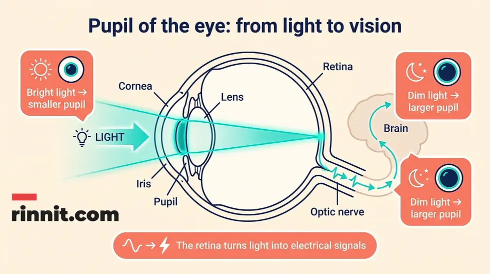
The pupil allows light to enter the eye. The lens focuses the light that passes through the pupil, and the light then reaches the retina at the back of the eye.
The retina changes light into electrical signals. The brain receives those signals and turns them into images. This sequence allows you to see.
The pupil is therefore an opening, not a colored structure. The iris surrounds it and adjusts its size to help regulate how much light enters.
Vision step | What happens |
Light enters | Light passes through the cornea and pupil |
Light is focused | The lens focuses light toward the back of the eye |
Light is converted | The retina turns light into electrical signals |
Signals are interpreted | The brain turns those signals into images |
Why do pupils get bigger and smaller?
In bright light, the pupil constricts and becomes smaller. In dim light, it dilates and becomes larger. This automatic reaction is called the pupillary light response.
Changing pupil size helps the eye respond to the available light. A professional may check this response by shining a light into the eye and observing whether the pupil changes size.
Light setting | Typical pupil response | Everyday meaning |
Bright light | Constriction, or a smaller pupil | Limits the amount of light entering the eye |
Dim light | Dilation, or a larger pupil | Allows more light to enter the eye |
Near focus | Pupils normally constrict when focusing on a nearby object | Helps coordinate near vision |
A person’s pupil response can vary with medications, eye conditions, nerve pathways and brain conditions. A response that seems unusual should be assessed in context.
Which muscles control the pupil?
Two muscles in the iris control how the pupil opens and closes.
The iris dilator muscle opens the pupil from the inside out. Its fibers extend outward from the center and can look a little like sun rays.
The iris sphincter is a circular muscle. It helps close the pupil and make the opening smaller.
Iris muscle | Main action |
Iris dilator muscle | Opens the pupil and makes it larger |
Iris sphincter | Closes the pupil and makes it smaller |
These muscles respond through nerve pathways that connect the eye with the brain.
How are the pupil and brain connected?
Nerves that affect the pupil connect to an afferent pathway and an efferent pathway.
The afferent pathway carries information from the eye to the brain. The efferent pathway carries signals from the brain back to the eye.
Because pupil reactions involve the eye, nerves and brain, an abnormal response can be a clue that a professional needs to evaluate more than the eye itself. A pupil finding alone does not establish a diagnosis.
Does the pupil have another role inside the eye?
The pupil also gives aqueous humor a pathway to the front of the eye. Aqueous humor is a fluid that nourishes the eyes.
The pupil’s best-known role is regulating the amount of light entering the eye, but its position also connects the back and front parts of the eye through the opening in the iris.
What conditions can affect pupil size or behavior?
A difference in pupil size or an unusual reaction to light can be an eye finding by itself or a sign of another condition.
Conditions associated with pupil size or behavior include Marcus Gunn pupil, anisocoria, polycoria, Argyll Robertson pupil, miosis, mydriasis and Adie syndrome.
Condition or finding | Plain-language meaning |
Marcus Gunn pupil | The pupils do not respond normally when a light is shined into the eyes |
Anisocoria | One pupil is larger than the other; it may be normal, temporary or related to a serious condition |
Polycoria | More than one working pupil is present in one eye |
Argyll Robertson pupil | Small pupils constrict when focusing on a nearby object but not normally in bright light |
Miosis | Very small or constricted pupils |
Mydriasis | Dilated pupils |
Adie syndrome | A pupil stays dilated or constricted and does not respond normally to light |
The same visible change can have different causes. A professional considers the timing, symptoms, medications, eye history and neurologic findings together.
What is anisocoria?
Anisocoria is the medical term for pupils that are different sizes.
Anisocoria can be normal or temporary in some people. It can also be caused by serious conditions. A new or changing difference in pupil size deserves professional attention, especially when it occurs with pain, vision changes, head injury or other neurologic symptoms.
The appearance of unequal pupils does not identify the cause. An eye-care professional can compare the pupils in different lighting and assess their response.
What are miosis and mydriasis?
Miosis is the medical term for very small or constricted pupils. Small pupils can happen after a stroke or after opioid use, among other reasons.
Mydriasis is the opposite condition: dilated pupils. A pupil may become larger in dim light, but persistent or unexpected dilation can have other causes.
Term | Meaning |
Miosis | Very small, constricted pupil |
Mydriasis | Dilated, enlarged pupil |
Anisocoria | Unequal pupil size |
These terms describe pupil appearance or behavior. They do not, by themselves, explain why the change occurred.
What is Marcus Gunn pupil?
Marcus Gunn pupil means that the pupils do not respond normally when an eye-care specialist shines a light into the eyes.
The finding is identified during an examination of the pupil response. It can prompt a professional to assess the eye and the nerve pathways involved in the light reaction.
What are Adie syndrome, Horner syndrome and third cranial nerve palsy?
Adie syndrome can cause a pupil to stay dilated and respond abnormally to changes in light. It can also cause a pupil to stay constricted rather than dilate.
Horner syndrome is a rare condition that affects the eye and face, usually on one side. It is also called oculosympathetic palsy. Palsy refers to nerve paralysis.
The third cranial nerve is the oculomotor nerve. It controls muscles that move the eye, constrict the pupil, focus the eyes and position the upper eyelid. Palsy of this nerve can affect several of these functions.
Condition | Pupil-related feature |
Adie syndrome | Pupil remains dilated or constricted and reacts abnormally to light |
Horner syndrome | A rare one-sided eye-and-face nerve condition that can affect the pupil |
Third cranial nerve palsy | Affects a nerve involved in pupil constriction, eye movement, focusing and upper-eyelid position |
These conditions require professional evaluation. Their names should not be used to self-diagnose from a photograph or a brief change in pupil size.
What other conditions can affect the pupils?
The pupil can change in connection with traumatic brain injury, cataracts, cataract surgery and headaches.
A traumatic brain injury occurs when the brain hits against the skull. A concussion is one of the most common types. One of the first things a professional may check after a brain injury is how the pupils look and respond.
Cataracts make the normally clear lenses of the eyes cloudy. Cataract surgery may affect pupil size, and pupil size may affect complications after surgery.
Cluster headaches often affect one side of the head and therefore one eye or pupil. One eye may water during an attack.
Related condition | Possible relationship to the pupil |
Traumatic brain injury | Pupil appearance and response may be checked during assessment |
Cataracts | Cloudy lenses and cataract surgery can affect or be affected by pupil size |
Cluster headaches | One-sided headache symptoms can involve one eye or pupil, sometimes with watering |
Stroke | Can be associated with very small pupils or other pupil changes |
Medication effects | Some medicines, including opioids, can change pupil size |
The pupil finding is one part of an evaluation. It does not identify the cause by itself.
What tests check pupil health?
An eye-care specialist may use a comprehensive eye exam to assess the pupils and the rest of the visual system.
During an exam, a professional may shine a light into each pupil to see whether it gets bigger or smaller. Both pupils should react to light, although the meaning of a response depends on the full clinical context.
Medicated eye drops such as pilocarpine or phenylephrine may be used to test pupil reaction. These drops should only be used under professional direction.
If the pupils are not working as expected, a professional may order blood tests or imaging tests to look for other diseases. These may include serious conditions such as stroke or brain injury.
Evaluation method | What it may assess |
Comprehensive eye exam | Pupil reaction, vision and the health of the eye |
Light response check | Whether each pupil changes size when illuminated |
Medicated eye drops | How the pupil reacts to specific medicines |
Blood tests | Possible medical causes when indicated |
Imaging tests | Possible eye, nerve or brain causes when indicated |
A clinician selects tests based on the person’s symptoms, history and examination findings.
How are pupil conditions treated?
Treatment depends on what is causing the pupil change.
If a medication affects the pupils, a professional may suggest stopping the medicine or substituting another one. Do not change a prescription without speaking with the prescriber.
Some conditions may require medication in the form of eye drops or topical preparations. Other medications may be pills, capsules, liquids, injections or intravenous treatments.
Some eye conditions may be treated with eye exercises or surgery. The appropriate plan depends on the underlying condition and the health of the eye, nerves and brain.
Treatment category | How it may be used |
Medication review | A prescriber may adjust a medication that affects the pupils |
Eye drops or topical treatment | May be used for some eye conditions |
Oral, injected or intravenous medication | May be used when a wider medical treatment is needed |
Eye exercises | May be recommended for some conditions |
Surgery | May be considered for selected eye conditions |
Do not use diagnostic eye drops or attempt to treat an abnormal pupil without professional guidance.
When should someone seek urgent care for a pupil change?
A sudden new change in pupil size or reaction can need urgent assessment, particularly after a head injury or when other symptoms are present.
Seek urgent medical evaluation for sudden unequal pupils, a new abnormal pupil after head trauma, sudden vision loss, severe eye pain, confusion, weakness, speech changes, a severe headache or a rapidly worsening eye or neurologic symptom. These signs can have different causes and do not establish one diagnosis.
Situation | Appropriate next step |
Sudden pupil change with neurologic symptoms | Seek urgent medical evaluation |
New pupil change after head injury | Seek urgent evaluation rather than waiting for it to resolve |
Sudden vision loss or severe eye pain | Seek urgent eye or medical care |
Long-standing, stable difference without other symptoms | Arrange a professional eye assessment to discuss it |
If someone is difficult to wake, has severe breathing problems or has signs of a life-threatening emergency, call local emergency services.

How can someone care for their eyes?
General eye and pupil health is supported by everyday habits, though they cannot prevent every eye condition.
Avoid smoking and seek help to quit if you use tobacco. Eat a healthy diet that fits your needs, stay active and drink enough water. Wear sunglasses and safety glasses for appropriate work, sports and outdoor conditions.
If you wear contact lenses, wash your hands before putting them in or taking them out. Follow the manufacturer’s or eye-care professional’s instructions for cleaning and wear time.
During computer use, screen time or extensive reading, many people use the 20-20-20 rule: look at something 20 feet away for 20 seconds about every 20 minutes. It is a widely suggested comfort habit, but a small controlled study did not find that scheduled 20-second breaks significantly changed eye-strain symptoms, so it should not be relied on as a treatment.
Ask an eye-care professional how often you need vision screenings or eye exams. People with glaucoma or diabetes may need more frequent visits.
Eye-care habit | Practical action |
Avoid tobacco | Do not smoke; seek help to quit if needed |
Protect the eyes | Use sunglasses and safety eyewear when appropriate |
Handle contacts carefully | Clean hands, proper cleaning and recommended wear time |
Rest during close work | Take regular breaks from screens or reading, for example with the 20-20-20 habit |
Support overall health | Eat a healthy diet, stay active and drink enough water |
Keep eye visits | Follow the exam schedule recommended for your eyes and health conditions |
What questions should you ask an eye-care professional about your pupils?
Questions about your pupils can cover whether size differences are new, whether they react normally to light, whether medicines could affect them and which warning signs need urgent care.
Is the difference in pupil size new, temporary or long-standing?
Do both pupils constrict and dilate normally in different lighting?
Could a medication, eye drop or recreational substance affect pupil size?
Do my symptoms suggest that the eye, a nerve pathway or the brain needs assessment?
Would a comprehensive eye exam, blood test or imaging be useful?
What warning signs should prompt urgent care?
If I wear contact lenses, what habits are safest for my eyes?
How often should I have an eye exam?
Do glaucoma, diabetes, cataracts or a history of head injury change my follow-up schedule?
Conclusion
The pupil is the opening in the center of the iris that lets light into the eye. The iris muscles adjust its size, making the pupil smaller in bright light and larger in dim light. The lens focuses incoming light onto the retina, and the retina sends electrical signals to the brain so images can be formed.
Pupil changes can be normal, temporary or associated with eye, nerve or brain conditions. Anisocoria, miosis, mydriasis and abnormal light responses describe findings, not complete diagnoses. Professional evaluation is especially important when a pupil change is sudden, follows head injury or occurs with vision loss, severe eye pain or neurologic symptoms.
Want a clearer next step for eye-health questions? Read more about eye health and talk with an eye-care professional about any pupil changes before making decisions.
Frequently Asked Questions
What is the pupil?
The pupil is the black opening in the center of the iris, the colored part of the eye. It lets light enter the eye.
Why does the pupil get smaller in bright light?
The iris muscles constrict the pupil in bright light. This response helps regulate how much light enters the eye.
Why does the pupil get bigger in the dark?
The pupil dilates in dim light so more light can enter the eye. This is part of the pupillary light response.
What is the pupillary light response?
The pupillary light response is the automatic change in pupil size in response to light. Pupils normally become smaller in bright light and larger in dim light.
What is the relationship between the pupil and iris?
The pupil is the opening in the middle of the iris. The iris is the colored part of the eye and contains the muscles that adjust pupil size.
What is anisocoria?
Anisocoria means one pupil is larger than the other. It may be normal or temporary, but it can also be related to a serious condition, especially when it is new or occurs with other symptoms.
What are miosis and mydriasis?
Miosis means very small or constricted pupils. Mydriasis means dilated or enlarged pupils.
What is Marcus Gunn pupil?
Marcus Gunn pupil means the pupils do not respond normally when a professional shines a light into the eyes. It is an examination finding that may require further assessment.
What is Adie syndrome?
Adie syndrome can cause a pupil to remain dilated or constricted and to respond abnormally to changes in light.
Can a head injury affect the pupils?
Yes. After a traumatic brain injury, a professional may check the pupils’ appearance and responses as part of the assessment. A new pupil change after head injury needs urgent evaluation.
Can medication change pupil size?
Yes. Some medications can affect the pupils. Opioids, for example, can be associated with small pupils. Do not stop or change a medication without speaking with the prescriber.
What tests may be used to evaluate the pupil?
A comprehensive eye exam may include checking the response to light. Depending on the findings, a professional may use medicated eye drops or order blood tests or imaging tests.
How are pupil conditions treated?
Treatment depends on the cause. Options can include medication review, eye drops, other medications, eye exercises or surgery. A professional should guide treatment.
When should someone seek urgent care for a pupil change?
Seek urgent evaluation for sudden unequal pupils, a new pupil change after head injury, sudden vision loss, severe eye pain, confusion, weakness, speech changes, a severe headache or a rapidly worsening eye or neurologic symptom.
How can someone protect their eyes?
Avoid tobacco, eat a healthy diet, stay active, wear appropriate sunglasses and safety eyewear, follow safe contact-lens habits, take regular breaks during screen work and keep regular eye exams as recommended.
Related Reading
PERRLA Eye Exam: What It Means and What Pupil Changes Can Show explains the quick pupil check used in eye and neurological exams.
References
Lam BL, Thompson HS, Corbett JJ. The prevalence of simple anisocoria. Am J Ophthalmol. 1987;104(1):69-73. PMID 3605282. https://doi.org/10.1016/0002-9394(87)90296-0
Kiani I, Parsaei M, Karimi H, et al. Prognostic role of quantitative pupillometry in traumatic brain injury: a scoping review. Neurol Sci. 2025;46(3):1169-1177. PMID 39663272. https://doi.org/10.1007/s10072-024-07869-y
Johnson S, Rosenfield M. 20-20-20 rule: are these numbers justified? Optom Vis Sci. 2023;100(1):52-56. PMID 36473088. https://doi.org/10.1097/OPX.0000000000001971
National Eye Institute. Get a Dilated Eye Exam. National Eye Institute. Last updated November 26, 2025. https://www.nei.nih.gov/eye-health-information/healthy-vision/finding-eye-doctor/get-dilated-eye-exam
Centers for Disease Control and Prevention. Concussion Basics. CDC. Accessed October 30, 2026. https://www.cdc.gov/heads-up/about/index.html
Disclaimer
This article is for general educational purposes and is not a substitute for professional medical advice, diagnosis or treatment. Always seek the advice of a qualified health provider with any questions you may have regarding a medical condition.
It explains pupil anatomy, the pupillary light response, iris muscles, the lens-retina-brain vision pathway, aqueous humor, pupil findings and general eye care. It does not diagnose anisocoria, miosis, mydriasis, Marcus Gunn pupil, Adie syndrome, Horner syndrome, third cranial nerve palsy, stroke, traumatic brain injury, glaucoma or another eye or neurologic condition. Do not use medicated eye drops, stop a medication or perform eye exercises without professional direction.



Comments