Stereotactic Breast Biopsy: Why It's Done, What to Expect, Recovery and Results

Updated: 2 days ago
Written and medically reviewed by Dr. Baraa Alnahhal, MD · Last reviewed: November 2026 · How we review
Quick Answer
A stereotactic breast biopsy uses mammography to guide a needle precisely to a suspicious spot that can be seen on imaging but not felt, such as a cluster of calcifications. It's done under local anesthesia and leaves a tiny marker clip. Fewer than 30% of biopsies find cancer, and results usually arrive within a few days to a week.
What the research says
Uncertain ("B3") results are upgraded about one time in five: a meta-analysis of 78 studies and 6,377 lesions found 19% of lesions of uncertain malignant potential found on core biopsy turned out to be cancer at surgery, with atypical ductal hyperplasia (ADH) and lobular neoplasia carrying the highest upgrade rates (Cullinane C et al., European Journal of Surgical Oncology, 2022) [2]
Surgery vs follow-up after ADH: among 6,458 ADH diagnoses on core biopsy, 29% were upgraded to cancer when excised surgically compared with 5% during imaging follow-up, which supports surgical excision for most ADH findings (Schiaffino S et al., Radiology, 2020) [3]
Key takeaways
Chosen when a mammogram shows calcifications, a small mass or distortion that ultrasound cannot see.
You lie face down (or sit upright) with the breast held gently between two plates; local anesthetic numbs the area.
A vacuum-assisted needle removes several small cores through one tiny nick; a marker clip is left in place.
The whole visit lasts about an hour; the sampling itself takes minutes.
Most results are benign; an uncertain result may lead to surgical removal because around one in five is upgraded.
What is a stereotactic breast biopsy?
"Stereotactic" means the target is located in three dimensions using two mammogram images taken from different angles, or a digital tomosynthesis sweep. A computer calculates the exact coordinates, and the needle is advanced to that point. It is the method of choice for abnormalities that show up only on mammography. Sources: [1]
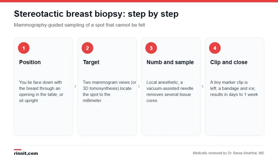
A stereotactic biopsy is guided by mammogram images and takes only minutes once the target is located.
Why is it done?
Your radiologist may recommend it after a screening mammogram shows: Sources: [1]
Suspicious or new microcalcifications.
A small mass or area of architectural distortion not visible on ultrasound.
A change in the breast compared with earlier mammograms.
An abnormality that needs tissue confirmation before surgery is planned.
How do I prepare?
Tell your provider about blood thinners, aspirin and supplements; you may be asked to pause them.
Do not use deodorant, powder or lotion on the breast or underarm on the day of the test.
Wear a two-piece outfit and a supportive bra to wear afterward.
Arrange a ride home if you take a sedative, although most people do not need one. Sources: [1]
What happens during the procedure?
You lie face down on a special table with the breast through an opening, or sit upright at a mammography unit.
The breast is compressed gently between two plates and images are taken to locate the target.
The skin is cleaned and numbed with local anesthetic.
A small nick is made and a hollow vacuum-assisted needle removes several tissue cores.
A tiny titanium or ceramic marker clip is placed so the site can be found again.
A final mammogram confirms the clip position; a bandage is applied.
You will feel pressure and may hear clicking from the device, but sharp pain is uncommon. Expect to be at the center for about an hour. Sources: [1]
Recovery and aftercare

Most people are back to routine activities the next day.
Apply ice for 20 minutes on, 20 minutes off for the first few hours to limit bruising.
Keep the dressing dry for 24 hours; showering is fine after that.
Avoid strenuous exercise and heavy lifting for 24–48 hours.
Mild soreness and a bruise are normal and fade within 1–2 weeks.
Call your provider for increasing swelling, bleeding that soaks the bandage, redness, warmth or fever. Sources: [1]
Understanding your results
A pathologist examines the cores, and the result is usually available within a few days to a week. Fewer than 30% of breast biopsies show cancer. Results fall into three groups: Sources: [1]
Result | What it means | Usual next step |
Benign | No cancer (for example fibroadenoma, cyst wall, benign calcifications) | Routine follow-up imaging |
Uncertain / high-risk (B3) | Atypical ductal hyperplasia, lobular neoplasia, flat epithelial atypia, radial scar, papillary lesion | Often surgical excision, sometimes close imaging follow-up |
Malignant | Ductal carcinoma in situ or invasive cancer | Referral to a breast cancer team for staging and treatment |
Research helps explain why uncertain results are often removed surgically. Across 78 studies, 19% of B3 lesions were upgraded to cancer at surgery, with ADH and lobular neoplasia upgraded most often, while flat epithelial atypia, radial scars and papillary lesions were benign more than 90% of the time. In a separate analysis of 6,458 ADH cases, 29% were upgraded when excised compared with 5% during imaging follow-up. Sources: [2, 3]

Pooled upgrade rates from two large meta-analyses of core biopsy results.
Risks and limitations
Bruising, bleeding or a small collection of blood (hematoma).
Infection at the needle site (rare).
Rarely, the target cannot be reached, for example in very thin breasts or lesions close to the chest wall.
Sampling error: a benign result that does not match the imaging may need a repeat biopsy or surgical biopsy. Sources: [1]
Conclusion
Stereotactic breast biopsy turns an abnormal mammogram into a clear tissue answer with a needle rather than an operation. Most results are benign. If yours is uncertain or malignant, the marker clip left during the biopsy helps your surgeon find the exact spot, and the research on upgrade rates helps your team decide between surgery and careful follow-up.
Keep learning: see our guides to breast biopsy types and to breast calcifications.
Frequently asked questions
Does a stereotactic breast biopsy hurt?
The skin is numbed with local anesthetic, so most people feel pressure and some pulling rather than pain. Soreness and bruising afterward are common and mild.
How long does it take?
Plan on about an hour at the imaging center. The actual sampling takes only a few minutes; most of the time is spent positioning and imaging.
Why do I need it instead of an ultrasound-guided biopsy?
Calcifications and some small distortions are visible only on mammography, so the mammogram itself is used to guide the needle.
What is the clip left in my breast?
A tiny titanium or ceramic marker that shows the biopsy site on future imaging. It is safe with MRI and airport scanners and does not need to be removed.
How accurate is a stereotactic biopsy?
Core biopsy is highly accurate, but a small number of uncertain results are upgraded to cancer at surgery, which is why some benign-appearing findings are still removed.
What does a B3 or "atypical" result mean?
It means the cells look abnormal but not clearly cancerous. About one in five of these is upgraded to cancer at surgery, so excision is often recommended.
When will I get my results?
Usually within a few days to one week. Your provider or the radiologist will call you or review them at a follow-up visit.
Can I drive home afterward?
Yes, unless you were given a sedative. Most people drive themselves and return to normal activities the next day.
Will it leave a scar?
The nick is only a few millimeters and usually heals with a barely visible mark.
What if the result is benign but my mammogram still looks suspicious?
Your radiologist checks that the result matches the imaging. If it does not, a repeat or surgical biopsy may be recommended.
Related Reading
Breast Biopsy: Types, What to Expect, Recovery and Results explains all biopsy approaches, including ultrasound- and MRI-guided options.
Mammogram: Types, How to Prepare, What Happens and Your Results covers the imaging that usually leads to a stereotactic biopsy.
Breast Calcifications: What They Are, What They Mean, and When to Follow Up the most common reason this biopsy is ordered.
Breast Cancer Screening: When to Start, How Often and Which Tests Are Used when to start screening and which tests are used.
Breast Cancer: Symptoms, Causes, and Treatment what happens if the biopsy finds cancer.
Core Needle Breast Biopsy: Procedure, Recovery and Results explains the needle biopsy used to check breast changes for cancer.
References
Cleveland Clinic. Stereotactic Breast Biopsy. Cleveland Clinic. Last updated October 4, 2022. https://my.clevelandclinic.org/health/diagnostics/24256-stereotactic-breast-biopsy
Cullinane C, Byrne J, Kelly L, et al. The positive predictive value of vacuum assisted biopsy (VAB) in predicting final histological diagnosis for breast lesions of uncertain malignancy (B3 lesions): a systematic review & meta-analysis. Eur J Surg Oncol. 2022;48(7):1464-1474. PMID 35491362. https://doi.org/10.1016/j.ejso.2022.04.005
Schiaffino S, Calabrese M, Melani EF, et al. Upgrade rate of percutaneously diagnosed pure atypical ductal hyperplasia: systematic review and meta-analysis of 6458 lesions. Radiology. 2020;294(1):76-86. PMID 31660803. https://doi.org/10.1148/radiol.2019190748
Medical disclaimer
This article is for general education only and does not replace advice from a qualified healthcare professional. Talk to your doctor about your own diagnosis and treatment, and seek emergency care right away for severe or sudden symptoms.



Comments