Loopogram: How It Checks Your Ileal Conduit After Bladder Removal

Updated: 3 days ago
Written and medically reviewed by Dr. Baraa Alnahhal, MD · Last reviewed: November 2026 · How we review
Quick Answer
A loopogram is an X-ray test that checks how well urine drains through an ileal conduit, a urine pathway made from small intestine after the bladder is removed. Contrast fluid is injected through a catheter in the stoma, and X-rays help find narrowing, leaks or blockages. The test takes 15 minutes. It isn't done during pregnancy.
What the research says
Stoma problems after an ileal conduit are common: a systematic review of 14 studies found external stoma and skin complications after ileal conduit diversion were reported in 15% to 65% of patients, including hernias, narrowing (stenosis), prolapse and skin irritation, and that these problems are often under-reported (Szymanski KM et al., Ostomy/Wound Management, 2010) [2]
An ileal conduit has fewer early complications than continent diversions: in 4,755 people who had their bladder removed for cancer, those who received a continent diversion had 1.4 times the odds of a complication and 1.7 times the odds of readmission within 30 days compared with an ileal conduit (Rezaee ME et al., Urology, 2022) [3]
Key takeaways
A loopogram checks urine flow through an ileal conduit.
Contrast is injected through the stoma, and X-rays are taken.
It helps find narrowing, leaks or blockages.
The test takes about 15 minutes.
Bring a new stoma bag and supplies.
What is a loopogram?
After bladder removal surgery, often for bladder cancer, a surgeon may create an ileal conduit. The ureters, the tubes that carry urine from the kidneys, are connected to a short piece of small intestine that opens on your belly as a stoma. Urine then drains into a bag, called a urostomy. A loopogram shows whether this pathway is working properly. A similar test can also check some colostomies or ileostomies. Sources: [1]
Why it's done

A loopogram checks the whole urinary pathway.
Check urinary tract function after urinary diversion surgery.
Look for narrowing (strictures) where the ureters join the conduit.
Find leaks or blockages.
Help guide treatment decisions. Sources: [1, 2]
How to prepare

Preparation is simple.
Wear comfortable clothes and little jewelry; you'll change into a gown.
Bring a new stoma bag and dressings.
You can usually eat, drink and take your medicines as usual.
Tell your provider if you might be pregnant; you may need a pregnancy test. Sources: [1]
What happens during the test
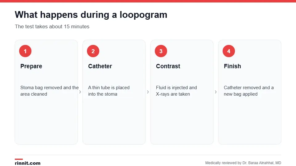
The test takes about 15 minutes.
You lie on the X-ray table, and your stoma bag is removed.
The area around the stoma is cleaned.
A thin catheter is placed into the stoma.
Contrast fluid is injected.
Several X-rays are taken, possibly in different positions.
The catheter is removed, and a new stoma bag is applied. Sources: [1]
A radiologist or radiology technician usually performs the test. Sources: [1]
Risks and recovery
Risks are minimal. There's a small chance of infection or an allergic reaction to the contrast. Most people return to normal activities right away. Sources: [1]
Results
A radiologist reviews the images and sends a report to your provider. If narrowing, a leak or another problem is found, your provider will discuss treatment options and next steps. Sources: [1, 2]
Conclusion
A loopogram is a short, low-risk X-ray test that makes sure your ileal conduit is draining urine as it should. Regular checks help catch problems early and protect your kidneys.
Keep learning: read about the urostomy and cystectomy.
Frequently asked questions
What is a loopogram?
An X-ray test of urine flow through an ileal conduit.
Who needs a loopogram?
People with an ileal conduit after bladder removal.
How long does it take?
About 15 minutes, with 30 to 60 minutes for the whole visit.
Does a loopogram hurt?
Most people feel only mild pressure when the catheter is placed.
Do I need to fast?
No, you can usually eat and drink as usual.
What should I bring?
A new stoma bag and dressings.
Can I have a loopogram while pregnant?
No, it shouldn't be done during pregnancy.
What are the risks?
A small risk of infection or contrast allergy.
Who performs it?
A radiologist or radiology technician.
What happens if a problem is found?
Your provider will discuss treatment options.
Related Reading
Urostomy: A Comprehensive Guide to Urinary Diversion Surgery living with the stoma bag an ileal conduit drains into.
Cystectomy (Bladder Removal Surgery): Types, Procedure, Risks and Recovery the surgery that leads to an ileal conduit.
Robotic Cystectomy: A Comprehensive Guide to Minimally Invasive Bladder Surgery a less invasive way to remove the bladder.
Understanding the Indiana Pouch: A Comprehensive Guide to Urinary Diversion another type of urinary diversion.
BCG Treatment for Bladder Cancer: How It Works, Schedule, Side Effects and Aftercare a treatment for early bladder cancer.
References
Cleveland Clinic. Loopogram: Purpose, Procedure, Risks, Results & Recovery. Cleveland Clinic. Last updated March 11, 2024. https://my.clevelandclinic.org/health/diagnostics/15335-loopogram
Szymanski KM, St-Cyr D, Alam T, et al. External stoma and peristomal complications following radical cystectomy and ileal conduit diversion: a systematic review. Ostomy/Wound Management. 2010;56(1):28-35. PMID 20093715. https://pubmed.ncbi.nlm.nih.gov/20093715/
Rezaee ME, Atwater BL, Bihrle W, et al. Ileal conduit versus continent urinary diversion in radical cystectomy: a retrospective cohort study of 30-day complications, readmissions, and mortality. Urology. 2022;170:139-145. PMID 36007686. https://doi.org/10.1016/j.urology.2022.08.020
Medical disclaimer
This article is for general education only and does not replace advice from a qualified healthcare professional. Talk to your doctor about your own diagnosis and treatment, and seek emergency care right away for severe or sudden symptoms.



Comments