Deep Tendon Reflexes (DTRs): How They're Tested, Graded and What Abnormal Reflexes Mean

Updated: 2 days ago
Written and medically reviewed by Dr. Baraa Alnahhal, MD · Last reviewed: November 2026 · How we review
Quick Answer
Deep tendon reflexes (DTRs) are automatic muscle responses that a provider tests by tapping a tendon with a rubber reflex hammer. Each is graded from 0 (absent) to 4+ (very brisk), with 2+ typical. Reflexes that are too weak, too strong or different on each side can point to problems in the nerves, spinal cord or brain.
What the research says
Reflexes help diagnose spinal cord compression: in a study of patients with suspected degenerative cervical myelopathy, a simple model using the patellar reflex, Hoffmann reflex, a 10-second grip-and-release test, age and sex distinguished people with the condition with excellent accuracy (AUC 0.956) (Funaba M et al., Journal of Bone and Joint Surgery, 2025) [2]
Reflexes help detect nerve damage: in kidney transplant recipients, a short clinical score combining numbness and tingling, pinprick testing and Achilles and patellar reflex testing identified polyneuropathy with high accuracy (AUC 0.90) (Nolte S et al., Journal of the Peripheral Nervous System, 2025) [3]
Key takeaways
Five reflexes are commonly tested: biceps, brachioradialis, triceps, patellar and Achilles.
Graded 0 to 4+; 2+ is typical, and 3+ can also be normal.
Weak or absent reflexes often suggest nerve or muscle problems; very brisk reflexes can suggest brain or spinal cord problems.
Comparing both sides is important, because people vary naturally.
The Jendrassik maneuver helps bring out leg reflexes that seem absent.
What are deep tendon reflexes?
A reflex is an automatic response your nervous system makes without conscious thought. When a tendon is tapped, it stretches the attached muscle. Sensors in the muscle send a signal to the spinal cord, which sends a signal straight back, making the muscle contract. This loop keeps muscles ready to react and helps with balance. Because each reflex runs through specific nerves and spinal cord levels, checking them helps providers pinpoint where a problem is. They're part of every neurological exam. Sources: [1]
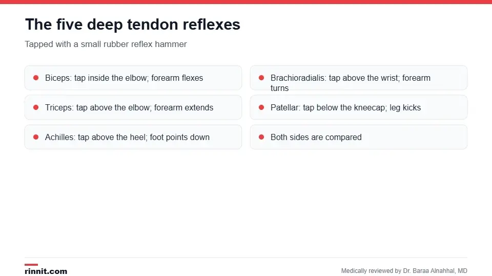
The five reflexes most often checked.
How are reflexes tested?
Reflex | Where the tap is | Normal response |
Biceps | Provider's thumb on the inside of the elbow | Forearm flexes |
Brachioradialis | Just above the wrist | Forearm flexes and turns; fingers may jerk |
Triceps | Just above the elbow at the back | Forearm extends |
Patellar (knee jerk) | Just below the kneecap | Lower leg kicks out |
Achilles (ankle jerk) | Achilles tendon above the heel | Foot points down |
You sit relaxed, and the provider uses a firm rubber reflex hammer during a routine office visit. Try not to think about the muscle being tested. Sources: [1]
The Jendrassik maneuver
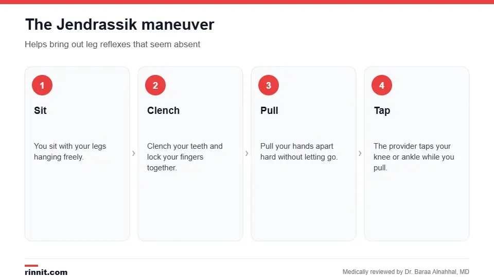
A simple trick to bring out leg reflexes.
If your knee or ankle reflex doesn't appear, your provider may ask you to sit with your legs hanging, clench your teeth, lock your fingers together and pull your hands apart while they tap. Your brain may unconsciously suppress the reflex when it expects the tap; the maneuver keeps it busy so the reflex shows normally. Sources: [1]
How reflexes are graded

Reflexes are graded from 0 to 4+.
Grade | Meaning |
0 | No reflex |
1+ | Reduced, less than expected |
2+ | Normal |
3+ | Brisker than average, can still be normal |
4+ | Very brisk, overactive, often with clonus (repeated rhythmic jerking) |
Reflex size varies naturally, so providers compare the same reflex on both sides and look at the overall pattern. Sources: [1]
What abnormal reflexes can mean
Weak or absent reflexes (hyporeflexia): often point to the peripheral nerves or muscles, as in peripheral neuropathy, diabetic neuropathy, Guillain-Barré syndrome, a pinched nerve root, spinal muscular atrophy or an underactive thyroid.
Very brisk reflexes (hyperreflexia): often point to the brain or spinal cord, as in multiple sclerosis, stroke, cervical myelopathy, a spinal cord injury or ALS.
Differences between sides: can help locate a problem on one side of the body. Sources: [1, 2, 3]
An abnormal result is a clue, not a diagnosis. Your provider may order further tests, such as nerve conduction studies, an MRI or blood tests. Sources: [1]
Conclusion
Reflex testing takes seconds but tells your provider a lot about how your nerves, spinal cord and brain are working. Most reflex differences are harmless, but combined with other findings, they help pinpoint problems early. Seek care if you notice new weakness, numbness or trouble moving.
Keep learning: read about the neurological exam and peripheral neuropathy.
Frequently asked questions
What are the five deep tendon reflexes?
Biceps, brachioradialis, triceps, patellar (knee) and Achilles (ankle).
What is a normal reflex grade?
2+, though 1+ and 3+ can be normal for some people.
What does a 4+ reflex mean?
Very brisk reflexes, often pointing to a brain or spinal cord problem.
What do absent reflexes mean?
They can suggest nerve or muscle problems, but some healthy people have weak reflexes.
Does reflex testing hurt?
No. The tap is quick and painless.
What is the Jendrassik maneuver?
Clenching your teeth and pulling locked hands apart to bring out leg reflexes.
Why are both sides compared?
Reflex strength varies between people, so differences between sides are more telling.
What is clonus?
Repeated rhythmic jerking after a tendon is stretched, often seen with very brisk reflexes.
Can thyroid problems affect reflexes?
Yes. An underactive thyroid can slow reflexes; an overactive thyroid can make them brisker.
What happens after an abnormal reflex test?
Your provider may order nerve tests, imaging or blood tests to find the cause.
Related Reading
Neurological Exam: What It Checks, What to Expect and What Happens Next the full exam that includes reflex testing.
Peripheral Neuropathy: Symptoms, Causes, and Treatment Options Explained nerve damage that can weaken reflexes.
What Is Cervical Myelopathy? Causes, Symptoms, Diagnosis and Treatment spinal cord compression that can make reflexes brisk.
Guillain-Barré Syndrome: When the Immune System Turns on Your Nerves, and How Treatment Helps You Recover a condition known for lost reflexes.
Amyotrophic Lateral Sclerosis (ALS): The Complete Guide a motor neuron disease that changes reflexes.
References
Cleveland Clinic. Deep Tendon Reflexes (DTRs): What They Are & How To Test. Cleveland Clinic. Last updated April 16, 2026. https://my.clevelandclinic.org/health/diagnostics/deep-tendon-reflexes-dtrs
Funaba M, Nakashima H, Tetreault L, et al. The utility of a prediction model using neurological examination findings for diagnosing degenerative cervical myelopathy. J Bone Joint Surg Am. 2025;107(16):1867-1875. PMID 40638695. https://doi.org/10.2106/JBJS.24.00098
Nolte S, Shehab NBN, Berger SP, et al. Polyneuropathy in kidney transplant recipients: accuracy of a new clinical diagnostic scoring system. J Peripher Nerv Syst. 2025;30(3):e70058. PMID 40915314. https://doi.org/10.1111/jns.70058
Medical disclaimer
This article is for general education only and does not replace advice from a qualified healthcare professional. Talk to your doctor about your own diagnosis and treatment, and seek emergency care right away for severe or sudden symptoms.



Comments