Accessory Navicular Bone: Symptoms, Types, Diagnosis and Treatment

Updated: 3 days ago
Medically reviewed by Dr. Baraa Alnahhal, MD · Last reviewed: October 2026
Quick Answer
An accessory navicular bone is an extra bone that forms near the navicular bone in the arch of the foot when extra cartilage hardens into bone during growth. Most cause no pain and need no treatment. It can be seen on an X-ray, and painful cases may be treated with supportive shoes, orthotics, immobilization, physical therapy or surgery [1].
What is an accessory navicular bone?
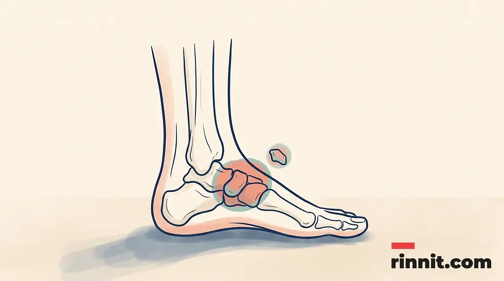
It is an extra bone that forms near the navicular bone in the arch of the foot, near the ankle. It develops when extra cartilage made during growth hardens into bone tissue. [1]
Does an accessory navicular always cause pain?
No. Most accessory navicular bones do not cause pain or other symptoms and do not need treatment. [1]
What is accessory navicular syndrome?
Accessory navicular syndrome is a symptomatic problem that can occur when an accessory navicular is large enough or positioned at an angle that places stress on the posterior tibial tendon or nearby bones. [1]
How is it diagnosed?
A healthcare provider can see an accessory navicular bone on an X-ray. The provider also considers the person’s symptoms and foot examination when discussing care. [1]
What treatment may help?
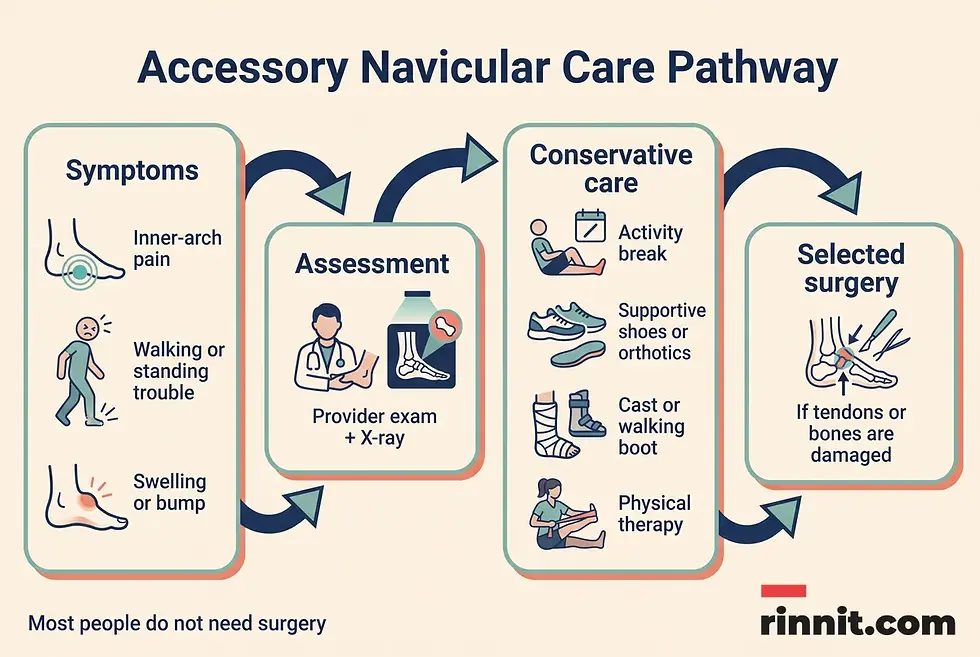
Depending on the symptoms, a provider may suggest activity changes, more supportive shoes, orthotics, pain medication, immobilization in a cast or walking boot, physical therapy or, in selected cases, surgery. [1]
What the research says
An accessory navicular is a common finding. A 2024 meta-analysis of 39 studies (11,015 people and 36,837 feet) estimated that about 17.5% of people and 12.6% of feet have one, and it was present in both feet in half of affected people. (Stolarz et al., Surg Radiol Anat, 2024) [2]
Only a minority of people develop pain. A 2023 review reports that about 10% of people with an accessory navicular have pain that affects walking and daily life. (Xiang et al., World J Clin Cases, 2023) [3]
What is an accessory navicular bone?
An accessory navicular bone is an extra bone in the foot that forms near the regular navicular bone. The navicular is a small bone between the ankle and the top of the foot’s arch. The terms os naviculare and os tibiale externum may also be used for an accessory navicular. [1]
Accessory navicular bones form during development. Bones begin as cartilage in infancy; if extra cartilage forms in the foot as the body develops, it may harden into an additional bone. Accessory naviculars can also be genetic, meaning a person may have a higher chance of having one if a biological parent has one. [1]
An accessory navicular is often an incidental finding: a person may have one without knowing it. Problems arise when the extra bone creates pressure on nearby tendons or bones, contributes to pain or increases the risk of certain injuries. [1]
Where is the accessory navicular located?
The accessory navicular forms near the actual navicular bone in the inner arch of the foot, near the ankle. Its relationship with the posterior tibial tendon is important because some types are embedded in or connected near that tendon. [1]
The posterior tibial tendon connects one of the calf muscles to bones on the inner side of the foot and ankle. If an accessory navicular is large or positioned in a way that repeatedly stretches or redirects this tendon, symptoms may develop. A tendon tear or rupture is possible but very rare. [1]
What are the three types of accessory navicular bone?
Healthcare providers classify accessory navicular bones into three types based on their size and where they form. Type II is the type most likely to cause symptoms. [1]
Type | Description | Symptom context |
Type I | Embedded in the posterior tibial tendon; approximately 2–3 millimeters and usually small. | Usually does not cause symptoms. |
Type II | Triangle- or heart-shaped; about 12 millimeters long; next to the regular navicular and connected to it by extra cartilage. | Most likely type to cause symptoms. |
Type III | Similar to Type II but not completely separate; an extra growth fused to the main navicular. | May be associated with symptoms when it creates pressure or tendon problems. |
The type describes the bone’s location and relationship with nearby structures. It does not, by itself, determine whether a person will have pain or which treatment is appropriate. [1]

What is accessory navicular syndrome?
Accessory navicular syndrome describes symptoms related to an accessory navicular bone. It may occur when the extra bone is large enough or forms at an angle that damages or places continuing stress on the posterior tibial tendon. [1]
The tendon may be stretched or forced to move in an unusual way. A tear or rupture can occur, but this is very rare. [1]
Symptoms
Possible symptoms include pain near the inside edge of the foot’s arch, difficulty walking or standing, trouble putting weight on the foot, limping, pain that worsens while wearing shoes, swelling, skin discoloration and a bump that can be seen or felt. [1]
These symptoms do not prove that an accessory navicular is the cause. Foot pain can have more than one possible explanation, so a healthcare provider should evaluate frequent or persistent symptoms. [1]
Why symptoms may appear
Accessory navicular syndrome can occur without a clear trigger, but symptoms may be more likely after a foot or ankle sprain. Overusing the feet and ankles for physical work, sports or demanding hobbies may also increase risk. Shoes that are too tight can add pressure over the extra bone and contribute to pain. [1]
Symptoms can develop at any age, but the syndrome usually affects teens. Rapid changes in bones during adolescence may increase the likelihood of problems near an accessory navicular bone. [1]
Possible symptom or trigger | What to know |
Inner-arch pain | Pain near the inside edge of the foot’s arch. |
Weight-bearing difficulty | Trouble walking, standing or putting weight on the foot. |
Shoe-related discomfort | Limping or pain that worsens in shoes. |
Local changes | Swelling, skin discoloration or a visible or touchable bump. |
Physical stress | A sprain, overuse from activity or tight shoes may contribute to symptoms. |
When should foot pain be assessed?
Consider arranging a healthcare assessment if foot pain occurs more often than not, interferes with walking or standing, worsens in shoes, or is accompanied by swelling, discoloration or a noticeable bump. Persistent pain does not have to be accepted as something to live with. [1]
A clinician can examine the foot, review the history of injury or overuse and use an X-ray to see whether an accessory navicular is present. Do not assume that every inner-arch bump or pain is caused by an accessory navicular bone. [1]
How is an accessory navicular bone diagnosed?
A healthcare provider can see an accessory navicular bone on an X-ray. The provider uses that finding together with the person’s symptoms and examination to decide whether the bone is causing a problem and what care may be appropriate. [1]
The presence of an accessory navicular does not automatically mean that treatment is needed. If the bone is not painful and does not cause other symptoms, treatment is not necessary. [1]
How is an accessory navicular bone treated?
If an accessory navicular never causes pain or other symptoms, treatment is not needed. If it causes symptoms, a healthcare provider may recommend conservative care first. The plan varies with the person’s symptoms and needs. [1]
Activity changes and the RICE method
A provider may suggest the RICE method. Taking a break from physical activities can reduce extra stress and potential damage in the foot. [1]
Shoes and orthotics
Changing shoes or choosing more supportive footwear may reduce pressure and improve comfort. A provider may also recommend orthotics, which can be custom-made or over-the-counter shoe inserts. [1]
Pain medication
Pain and swelling may be managed with over-the-counter NSAIDs or acetaminophen. A provider may offer a cortisone shot when symptoms are more severe and other treatments have not worked. Ask a clinician or pharmacist whether a medication is appropriate for you, particularly if you have other health conditions or take other medicines. [1]
Immobilization
A provider may recommend a cast or walking boot for a few weeks. Immobilization supports the foot and takes pressure off it while it heals. [1]
Physical therapy
Physical therapy can strengthen the muscles in the legs and feet and help reduce extra pressure on the bones. A physical therapist can prescribe customized exercises and stretches based on the person’s needs. [1]
Surgery
Most people do not need surgery. A provider may suggest surgery if the accessory navicular damages tendons or other bones. The operation may involve removing the extra bone and repairing damaged tendons. [1]
Care option | What it may involve |
Activity modification | A break from activities that add stress to the foot. |
Footwear or orthotics | More supportive shoes or custom-made or over-the-counter inserts. |
Symptom relief | Over-the-counter NSAIDs or acetaminophen; a provider may consider a cortisone shot in selected cases. |
Immobilization | A cast or walking boot for a few weeks. |
Physical therapy | Customized exercises and stretches to strengthen the legs and feet. |
Surgery | Removal of the extra bone and repair of damaged tendons when needed. |
How can people live well with an accessory navicular bone?
Having an accessory navicular does not automatically mean that a person will have ongoing problems. Most people never experience symptoms. When symptoms do occur, they can usually be managed with appropriate care. [1]
A provider can explain what an X-ray shows and whether the bone is likely contributing to symptoms. Ask for guidance about activity, footwear, pain management and rehabilitation rather than trying to match your symptoms to a treatment plan online. [1]
What questions should you ask a healthcare provider about an accessory navicular bone?
Helpful questions about an accessory navicular bone include whether it explains your symptoms, whether you need an X-ray, which activities to pause and whether shoes, orthotics or physical therapy would help.
Question | Why it may help |
Could my symptoms be related to an accessory navicular bone? | It starts a focused discussion without assuming the diagnosis. |
Do I need an X-ray? | The provider can explain whether imaging would clarify the anatomy. |
Which activities should I pause? | Activity needs vary with symptoms and healing. |
Would different shoes or orthotics help? | A clinician can discuss support and pressure reduction. |
Should I use pain medication? | The provider or pharmacist can review safety and appropriateness. |
Would physical therapy be useful? | Therapy may be customized to the muscles and foot mechanics involved. |
When would surgery be considered? | Most people do not need surgery, but tendon or bone damage may change the discussion. |
Conclusion
An accessory navicular bone is an extra bone near the navicular in the inner arch of the foot. Most people never have symptoms, but an accessory navicular can sometimes place pressure on nearby bones or the posterior tibial tendon. [1]
Accessory navicular syndrome may cause inner-arch pain, difficulty bearing weight, shoe-related discomfort, swelling, skin discoloration or a visible bump. Symptoms can appear after a sprain, overuse or pressure from tight shoes, and the syndrome usually affects teens during periods of rapid bone change. [1]
Next step: If foot pain is frequent, affects walking or standing, or comes with swelling, discoloration or a noticeable bump, arrange an assessment with a healthcare provider. Ask whether an X-ray and a conservative care plan could help clarify the next step.
FAQ
Is an accessory navicular bone dangerous?
Most accessory navicular bones do not cause symptoms and never require treatment. In rare cases, ongoing stress on the posterior tibial tendon can contribute to a tear or rupture. [1]
Can an accessory navicular bone cause inner-foot pain?
Yes. Accessory navicular syndrome can cause pain near the inside edge of the foot’s arch, along with difficulty walking, standing or putting weight on the foot. [1]
Is accessory navicular syndrome common in teenagers?
Clinical guidance says the syndrome usually affects teens. Rapid bone changes during adolescence may increase the likelihood of problems near an accessory navicular bone. [1]
Can tight shoes make symptoms worse?
Yes. Shoes that are too tight can place extra pressure on the accessory navicular and contribute to pain or other symptoms. [1]
What is Type II accessory navicular?
Type II is a triangle- or heart-shaped bone about 12 millimeters long. It sits next to the regular navicular and connects to it with extra cartilage. It is the type most likely to cause symptoms. [1]
Do all accessory navicular bones need treatment?
No. If the bone does not cause pain or other symptoms, treatment is not needed. [1]
What can I do for accessory navicular pain?
A healthcare provider may suggest a break from aggravating activity, more supportive shoes, orthotics, pain medication, immobilization, physical therapy or, in selected cases, surgery. The right approach depends on the person’s needs. [1]
Is surgery always needed?
No. Most people do not need surgery. A provider may consider it if the accessory navicular damages tendons or other bones. [1]
Can an X-ray show an accessory navicular bone?
Yes. A provider can see an accessory navicular on an X-ray. [1]
How common is an accessory navicular bone?
It is fairly common. A large review estimated that about 17.5% of people have one, and most never have symptoms.
External Links
Related Reading
Flat Feet: When You Need Treatment — and When You Don't explains fallen arches, which can occur with accessory navicular problems.
Orthotics: Types, Uses and How Custom Shoe Inserts Work describes shoe inserts that support the arch and ease foot pain.
Foot X-Ray: What It Shows, Why It's Done, What to Expect and Results explains the imaging test used to spot an accessory navicular bone.
Understanding High Arch Feet: Causes, Symptoms, and Support covers another arch shape that changes how the foot bears weight.
References
Cleveland Clinic. Accessory Navicular Bone: What It Is, Symptoms & Treatment. Cleveland Clinic. Last updated April 7, 2026. https://my.clevelandclinic.org/health/body/accessory-navicular-bone
Stolarz K, et al. The prevalence and anatomy of accessory navicular bone: a meta-analysis. Surg Radiol Anat. 2024;46(10):1731-1743. PMID 39136746. https://doi.org/10.1007/s00276-024-03459-x
Xiang F, et al. Accessory navicular in children. World J Clin Cases. 2023;11(35):8256-8262. PMID 38130606. https://doi.org/10.12998/wjcc.v11.i35.8256
Disclaimer
This article is for educational purposes only and is not a diagnosis or individualized medical advice. Always consult a qualified healthcare provider about your own health, and seek prompt care for new or worsening symptoms.



Comments