What Is a Hemangioma? Symptoms, Treatment & When to Worry

Updated: 2 days ago
Medically reviewed by Dr. Baraa Alnahhal, MD · Last reviewed: September 2026
TL;DR: A hemangioma (infantile hemangioma), sometimes called a "strawberry birthmark," is a bright red birthmark made of a dense clump of extra blood vessels in the skin. It usually appears at birth or within the first month of life, most often on the face, scalp, chest, or back. About 1 in 20 children is born with one. Most hemangiomas need no treatment — they grow during the first year, then rest and slowly fade, with little trace by age 10. Treatment is considered only when the mark affects vision, breathing, hearing, or feeding, or sits in a cosmetically sensitive spot. A provider can usually diagnose a hemangioma just by looking at it.
Quick Answer
What it is — a bright red birthmark made of extra blood vessels in the skin — often called a "strawberry birthmark."
When it appears — usually at birth or in the first month of life, most often on the face, scalp, chest, or back.
How common — about 1 in 20 children is born with one — it is the most common "tumor" of infancy, meaning growth, not cancer.
The timeline — it grows fastest during the first year, then rests and slowly fades; most are gone by age 10.
Treatment — generally not needed because it fades on its own.
See a doctor if — it bleeds, forms a sore, looks infected, or affects vision, breathing, hearing, or bathroom ability.
Options when treatment is needed — beta blocker gels or liquids, corticosteroids, or laser surgery.
What Is a Hemangioma (Infantile Hemangioma)?

A hemangioma (pronounced he-man-jee-O-muh), also known as an infantile hemangioma or hemangioma of infancy, is a bright red birthmark. It looks like a rubbery bump or a flat red patch and is made up of extra blood vessels that cluster together in the skin. The mark shows up at birth or within the first month of life.
The word "tumor" appears in the medical literature because a hemangioma is the most common tumor of infancy — but here, tumor simply means a growth. It is not cancer.
Hemangiomas appear most often on the face, scalp, chest, or back — though they can form anywhere on the skin. They are common: roughly 1 in 20 children is born with one, and the number of babies born with this birthmark has been rising steadily over the past 30 years.
How Does a Hemangioma Develop and Fade? (The Timeline)
Most infantile hemangiomas follow a predictable life cycle. Understanding this timeline explains why doctors so often recommend simply watching and waiting.
Appearance (at birth or first month) — starts as a flat red mark, usually on the face, scalp, chest, or back.
Rapid growth (first year) — the mark may grow quickly into a spongy, rubbery bump that sticks out from the skin.
Rest phase (after first year) — growth stops; the mark holds steady.
Fading (years 1–5) — the mark begins to slowly go away.
Resolution (by age 5–10) — many are gone by age 5; most by age 10, with at most a faint discoloration or raised trace.
Most children have only one mark, though some have more than one. After a hemangioma goes away, the skin underneath may be slightly discolored or raised, but there is usually little trace of it.
What Are the Signs of a Hemangioma?
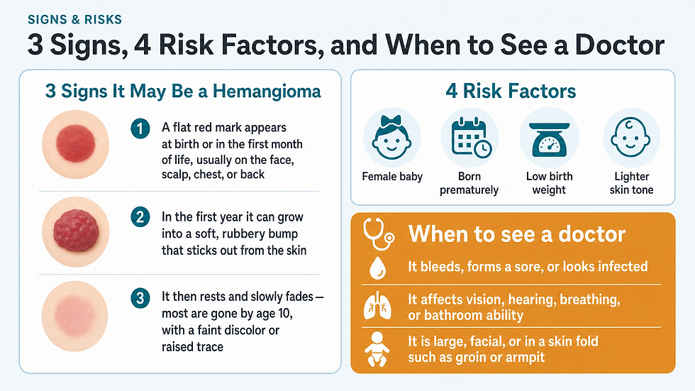
A hemangioma may be visible at birth, but it more often appears during the first month of life. The key signs:
A flat red mark appears at birth or soon after, most often on the face, scalp, chest, or back.
Rapid growth in the first year into a spongy, rubbery-looking bump that sticks out from the skin.
Slow fading afterward — many go away by age 5, and most by age 10, sometimes leaving slightly discolored or raised skin.
The fastest period of growth is the first 8 weeks of life. This matters for higher-risk marks, because acting within the window of rapid growth tends to leave a child with the best long-term outcome. For children whose hemangioma might be higher-risk, specialist evaluation around one month of life is ideal.
What Causes a Hemangioma?
A hemangioma is made up of extra blood vessels that group together into a dense clump. What causes those vessels to clump in the first place is not known. No cause has been identified — and nothing you did during pregnancy causes it.
What Increases the Risk?
Hemangiomas occur more often in babies who are female, white, born prematurely, or who have a low birth weight. Firstborn babies also carry somewhat higher odds, though many children with no risk factors — such as boys born at term with normal birth weight — develop them too.
Female sex — higher likelihood.
Lighter skin tone — higher likelihood.
Premature birth — higher likelihood.
Low birth weight — higher likelihood.
Firstborn child — slightly higher likelihood.
What Are the Possible Complications?
Most hemangiomas are harmless, but some need attention. A hemangioma can break down and develop a sore, which can lead to pain, bleeding, scarring, or infection.
Depending on its location, a hemangioma may also affect important bodily functions — vision, breathing, hearing, or the ability to go to the bathroom — though this is rare.
Certain patterns flag a mark as higher-risk and worth prompt specialist evaluation:
On the eyelid — can impair vision.
Involving the ear — can affect hearing.
Involving the mouth or lip — can impact feeding.
Large facial mark — can signal an associated condition called PHACE syndrome.
Multiple marks — can hint at internal hemangiomas, such as in the liver.
Large size, head/neck location, or skin folds (groin, armpit) — greater chance of bleeding, ulceration, or cosmetic concern.
When Should You See a Doctor?
Your child's health care provider will check any hemangioma during routine visits. Still, reach out between visits if the mark bleeds, forms a sore, or looks infected.
Seek medical care right away if the condition causes problems with an important bodily function — your child's vision, breathing, hearing, or ability to go to the bathroom. A good rule of thumb: any mark that is large, near the head or neck, in a skin fold, or at risk of significant cosmetic change deserves evaluation by someone who specializes in infantile hemangiomas.
How Is a Hemangioma Diagnosed?
In most cases, a health care provider can diagnose a hemangioma just by looking at it. Tests typically are not needed. No blood work, imaging, or biopsy is required for a typical skin hemangioma.
How Is a Hemangioma Treated?
Treating hemangiomas generally is not necessary, because they go away on their own over time. The decision to treat usually comes down to function or location: treatment is considered when the mark causes problems with vision, breathing, hearing, or feeding, or when it sits in a cosmetically sensitive area.
Beta blocker gel (timolol) — applied directly to the affected skin; best for small hemangiomas.
Beta blocker liquid (propranolol) — taken by mouth for hemangiomas that need stronger treatment; some go away with it. Side effects can include high blood sugar, low blood pressure, and wheezing.
Corticosteroids — given as a shot or applied to the skin when beta blockers do not work; side effects can include poor growth and thinning of the skin.
Laser surgery — used for a small, thin hemangioma, or sores on a hemangioma.
Beta blocker treatment typically continues until about 1 to 2 years of age. If you are considering treatment, talk it through with your child's health care provider — remember that most infantile hemangiomas go away on their own, and treatments can have side effects.

Living With a Hemangioma: What Parents Can Expect
For the large majority of families, the care plan is simple: watch and wait. Your provider will check the mark at routine visits. You can keep an eye on it at home by noting its size, color, and whether the surface stays intact. Photographing the mark every month or two gives your provider a useful record of how it is changing.
The emotional side matters too. A visible mark on a baby's face can worry parents, and it is completely reasonable to ask about treatment even when function is not affected. For marks in cosmetically sensitive areas, treatment options exist precisely because appearance matters to families — your provider can weigh the trade-offs with you.
Children with higher-risk marks may work with a team that includes pediatric ear, nose and throat doctors, eye doctors, pediatric plastic surgeons, neurologists, and pediatric radiologists. If your child's mark fits a higher-risk pattern, ask for a referral to a specialist who focuses on infantile hemangiomas.
Preparing for Your Appointment
Before your visit, note when the mark first appeared, how it has changed, and whether it has ever bled or formed a sore. Bring any photos you have taken over time. List any medications your child takes.
Questions to ask your health care provider:
Could this mark affect my child's vision, breathing, hearing, or feeding?
Is this mark in a higher-risk location or pattern?
Do we need treatment now, or is watching and waiting the right plan?
If we treat, which option fits my child's mark — gel, oral medicine, steroids, or laser?
What side effects should I watch for with this treatment?
How long would treatment last, and what happens after it stops?
What your provider will likely ask:
When did the mark first appear?
How quickly has it grown or changed?
Has it ever bled, formed a sore, or looked infected?
Does your child have more than one mark?
Was your child born prematurely or at a low birth weight?
Conclusion and Next Steps
A hemangioma is a common, bright red birthmark made of extra blood vessels — and in most cases, it is a chapter of infancy that closes on its own. The mark typically grows through the first year, rests, and slowly fades, with little trace by age 10. Tests are rarely needed, and treatment is usually not either.
The exceptions deserve attention. If the mark bleeds, sores, looks infected, or sits where it could affect vision, breathing, hearing, or feeding, contact your child's health care provider. When treatment is warranted, effective options exist — beta blocker gels and liquids, corticosteroids, and laser surgery — all best guided by a provider experienced with infantile hemangiomas.
Your next step: At your child's next routine visit, point out the mark and ask the questions in this guide. If your baby's hemangioma is large, facial, or near the eyes, ears, mouth, or skin folds, request an early evaluation — ideally within the first month of life, when the fastest growth happens and treatment works best.
Frequently Asked Questions
What does a hemangioma look like on a baby?
A hemangioma looks like a bright red birthmark. It starts as a flat red mark and often grows during the first year into a soft, spongy, rubbery bump that sticks out from the skin. It most often appears on the face, scalp, chest, or back.
Is a hemangioma a tumor? Is it cancer?
In medical language, a hemangioma is called the most common "tumor" of infancy — but tumor here means growth, not cancer. It is a benign clump of extra blood vessels, not a malignancy.
When does a strawberry birthmark go away?
Many hemangiomas go away by age 5, and most by age 10. After it fades, the skin may be slightly discolored or raised, but there is usually little trace of it.
Does a hemangioma need treatment?
Usually not. Most hemangiomas fade on their own without treatment. Treatment is considered when the mark causes problems with vision, breathing, hearing, or feeding, or when it sits in a cosmetically sensitive area.
How do you treat an infantile hemangioma?
When treatment is needed, options include a beta blocker gel (timolol) applied to the skin, an oral beta blocker liquid (propranolol) taken until about age 1–2, corticosteroids if beta blockers do not work, or laser surgery for small thin marks or sores.
What are the side effects of hemangioma treatment?
Oral propranolol can cause high blood sugar, low blood pressure, and wheezing. Corticosteroids can cause poor growth and thinning of the skin. Beta blocker gel applied to the skin is used for smaller marks. Discuss risks with your provider.
Can a hemangioma cause problems with vision or breathing?
It can, though rarely. Depending on location, a hemangioma may affect vision (eyelid), breathing, hearing (ear), feeding (mouth or lip), or bathroom ability. Any mark in these areas should be evaluated promptly.
How is a hemangioma diagnosed?
In most cases, a health care provider can diagnose a hemangioma just by looking at it. Tests typically are not needed.
References
Health information, not medical advice. This article is for general education and is not a substitute for professional diagnosis or treatment. Always consult a qualified healthcare provider about your own health, and seek emergency care for urgent symptoms.
Comments