Venogram (Venography): Why It's Done, What to Expect and Risks

Updated: 2 days ago
Written and medically reviewed by Dr. Baraa Alnahhal, MD · Last reviewed: November 2026 · How we review
Quick Answer: What is a venogram?
A venogram (venography) is an X-ray test that uses contrast dye and live imaging to show blood flowing through the veins, most often in the legs, pelvis and upper arms. Providers use it to find blood clots such as deep vein thrombosis when ultrasound can't give a clear answer. It takes 30 to 90 minutes, and you stay awake. [1]
What the research says
Ultrasound is now the usual first test, with venography as the reference. In a meta-analysis of 100 study groups that compared ultrasound with contrast venography in people with suspected DVT, ultrasound found 94.2% of clots in the thigh veins but only 63.5% in the calf veins, with 93.8% specificity. (Goodacre et al., BMC Medical Imaging, 2005) [2]
Inside-the-vein ultrasound can add to venography before stenting. In the VIDIO trial of 100 people with symptomatic pelvic vein narrowing (68 stented), intravascular ultrasound measurements predicted who improved after stenting, while venogram measurements did not. (Gagne et al., J Vasc Surg Venous Lymphat Disord, 2018) [3]
Key takeaways
A venogram shows blood moving through your veins using contrast dye and fluoroscopy (live X-ray). [1]
It is used when ultrasound is inconclusive, to diagnose pelvic vein problems, before some vein surgeries and during clot treatments. [1]
It is an invasive test: a needle is placed in a vein and you get a small dose of radiation. [1]
The test usually takes 30 to 90 minutes; drinking plenty of water for 24 hours afterward helps clear the dye. [1]
Main risks include reactions to the contrast dye, kidney injury and, rarely, a new blood clot. [1]
What is a venogram?
A venogram is an imaging test that shows how blood flows through your veins. Its main job is to help providers diagnose and manage vein conditions, including blood clots. [1]
Strictly speaking, "venography" is the procedure and "venogram" is the set of images it produces, but most people use "venogram" for both. [1]
Most vein clots are diagnosed with vascular ultrasound, which is noninvasive. A venogram is the alternative when ultrasound isn't possible or doesn't give enough detail, and it's also used to plan or guide treatment of vein disease. [1] [2]
Providers most often use venograms to look at veins in your: [1]
Legs
Pelvis
Upper arms
How does a venogram work?
Veins don't normally show up on an X-ray, so your provider injects a contrast dye into a vein. As the dye travels, it makes the vein visible and reveals clots or other flow problems. [1]
A single X-ray captures only one moment, but blood flow is constantly moving. So venograms use fluoroscopy, which fires short pulses of X-rays to create continuous, real-time pictures, much like many still frames joined into an animation. [1]
Why would you need a venogram?
Your provider may recommend a venogram to: [1]
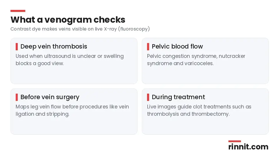
Reason | Examples |
Confirm or rule out a deep vein clot | Deep vein thrombosis (DVT) when ultrasound is unclear or swelling blocks a good view |
Check pelvic blood flow | Pelvic congestion syndrome, nutcracker syndrome and varicoceles |
Plan surgery | Mapping leg veins before procedures such as vein ligation and stripping |
Guide treatment in real time | Thrombolytic therapy and thrombectomy to dissolve or remove clots |
Sources: [1]
Venogram vs. angiogram
Both tests use X-rays and contrast dye to show blood moving through vessels. A venogram looks only at veins, while an angiogram can look at arteries or veins and is the gold standard for finding blocked arteries, sometimes with treatment such as angioplasty at the same time. Venograms are used less often for diagnosis because ultrasound usually finds vein blockages first. [1]
How do you prepare for a venogram?
Your provider will give you instructions. You may need to fast (only water) for a few hours, and you may be told to pause some medicines. [1]
Tell your provider about: [1]
All the medicines you take
Any medical conditions and allergies
Any past allergic reaction to contrast dye
Kidney disease or reduced kidney function, since some dyes can affect the kidneys
Any recent illness
Pregnancy or a chance you could be pregnant
On the day, remove jewelry and metal objects and wear loose, comfortable clothing; you may change into a gown. [1]
What happens during a venogram?
You are usually awake, though you may be offered a sedative to help you relax. A radiologic technologist takes you through these steps: [1]
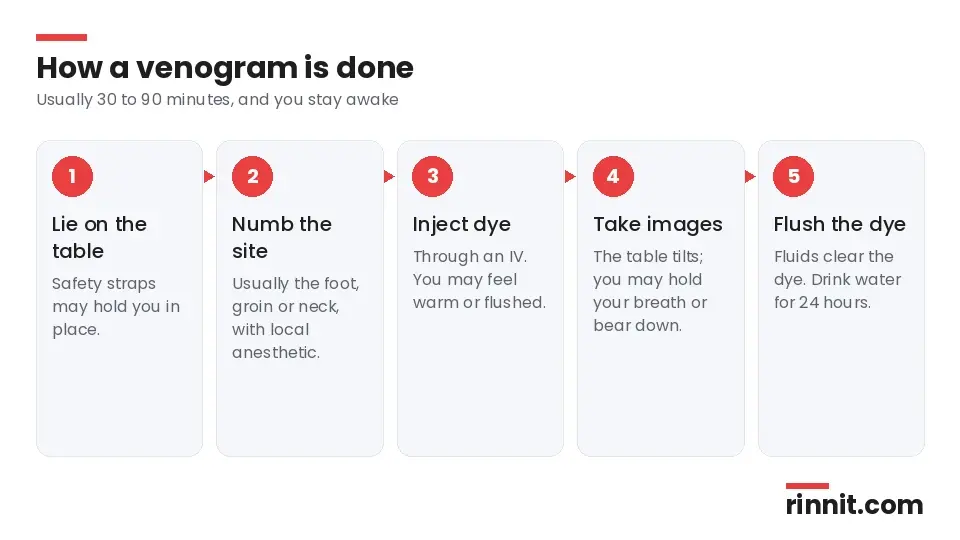
You lie on an X-ray table, sometimes with safety straps to keep you secure.
The team picks the access site, usually your foot, groin or neck, depending on which veins need to be seen.
A quick injection of local anesthetic numbs the site. It may feel like a pinch.
Contrast dye goes into your vein through an IV. You may feel pressure, and a warm or flushed feeling as the dye moves, which passes when the test ends.
The table may be tilted or moved to get different views. Tell the team if you feel uncomfortable.
You may be asked to hold your breath, stay very still or bear down (the Valsalva maneuver) to sharpen the images.
Fluids are run through the IV to flush out the dye.
If the venogram is guiding a treatment such as clot-dissolving therapy, your care team will add the steps needed for that treatment. [1]
What happens after a venogram?
You'll be watched for a short time for allergic reactions or other problems before you go home. You may be asked to drink plenty of water for the next 24 hours to help flush the dye from your body. [1]
What are the risks of a venogram?
For most people the benefits outweigh the risks, but a venogram may be too risky if you have certain conditions, such as severe kidney disease. Possible risks include: [1]
A small dose of radiation
Pain, bruising, bleeding or infection where the needle went in
An allergic reaction to the contrast dye
Kidney damage
Deep vein thrombosis (rare)
Contrast dye side effects
Brief nausea or a flushing feeling during the injection is common and passes quickly. Rarely, a reaction starts hours or days later. [1]

Severity | Possible symptoms |
Mild | Nausea and vomiting, headache, itchy skin, flushing, mild rash or hives |
Moderate | A rash or hives that gets worse, wheezing, abnormal heart rhythm, blood pressure that is too high or too low |
Severe | Trouble breathing, throat swelling, very low blood pressure |
Sources: [1]
Call your provider right away about any delayed reaction. Severe symptoms can be a sign of anaphylaxis, so seek emergency care immediately. [1]
What do venogram results mean?
A radiologist reviews the images and sends a report to the provider who ordered the test. Your provider will explain what the images show, for example a clot in a deep leg vein, and recommend treatment based on the findings. [1]
When a venogram is used to guide surgery, your provider uses the live images during the procedure and may not discuss separate results with you, but they will tell you if any problems come up. [1]
When should you call your provider?
Contact your provider if you: [1]
Have questions about why you need the test, how to prepare or what to expect
Notice side effects in the hours or days after the test
Have questions about your results or your next treatment steps
Conclusion
A venogram gives a live, detailed view of blood moving through your veins. Ultrasound is usually the first test for vein clots, but a venogram fills the gap when ultrasound falls short and helps guide treatments for clots and pelvic vein problems. [1] [2]
Share your full medical history, especially allergies and kidney problems, before the test, and get emergency help for any breathing trouble or throat swelling afterward.
Keep learning: Read our guide to deep vein thrombosis or our article on fluoroscopy. For more background, see RadiologyInfo: Venography.
FAQ
What is a venogram?
An X-ray test that uses contrast dye and live imaging to show blood flowing through your veins. [1]
Why would I need a venogram?
To check for blood clots when ultrasound is unclear, to look at pelvic vein problems, to plan vein surgery or to guide clot treatment. [1]
Is a venogram painful?
Most people feel only mild discomfort, such as a pinch from the numbing injection and warmth as the dye flows. [1]
Are you awake for a venogram?
Yes, you're usually awake, though you may get a sedative to help you relax. [1]
How long does a venogram take?
Usually 30 to 90 minutes. [1]
How do I prepare for a venogram?
You may need to fast for a few hours and pause some medicines; tell your provider about allergies, kidney problems and pregnancy. [1]
What are the risks of a venogram?
Radiation exposure, bruising or infection at the needle site, contrast dye reactions, kidney damage and, rarely, DVT. [1]
What should I do after a venogram?
Drink plenty of water for 24 hours to flush out the dye and report any delayed reaction to your provider. [1]
What is the difference between a venogram and an angiogram?
A venogram looks only at veins, while an angiogram can look at arteries or veins. [1]
Can I have a venogram if I'm pregnant?
It may not be safe during pregnancy, so tell your provider; they will decide whether the test is appropriate. [1]
Related Reading
Deep Vein Thrombosis (DVT): Symptoms, Causes, Diagnosis & Treatment — Complete Guide explains the leg vein clot a venogram can confirm when ultrasound is unclear.
What Are Varicose Veins? A Plain-Language Guide to the Bulging, Twisted Veins That Most Often Show Up on the Legs covers the vein problem that sometimes needs vein mapping before surgery.
Understanding May-Thurner Syndrome: Causes, Symptoms, and Care explains a pelvic vein compression condition where vein imaging guides care.
Thrombectomy: How Doctors Remove a Blood Clot, Risks, and Recovery describes a clot-removal procedure that venography can guide in real time.
References
Cleveland Clinic. Venogram: Procedure Details & Recovery. Cleveland Clinic. Last updated April 26, 2023. https://my.clevelandclinic.org/health/diagnostics/24929-venogram
Goodacre S, Sampson F, Thomas S, et al. Systematic review and meta-analysis of the diagnostic accuracy of ultrasonography for deep vein thrombosis. BMC Med Imaging. 2005;5:6. PMID 16202135. https://doi.org/10.1186/1471-2342-5-6
Gagne PJ, Gasparis A, Black S, et al. Analysis of threshold stenosis by multiplanar venogram and intravascular ultrasound examination for predicting clinical improvement after iliofemoral vein stenting in the VIDIO trial. J Vasc Surg Venous Lymphat Disord. 2018;6(1):48-56.e1. PMID 29033314. https://doi.org/10.1016/j.jvsv.2017.07.009
Medical disclaimer
This article is for general education only and does not replace advice from a qualified healthcare professional. Talk to your doctor about your own diagnosis and treatment, and seek emergency care right away for severe or sudden symptoms.



Comments