Understanding Tracheobronchomalacia (TBM)

Updated: 3 days ago
Medically reviewed by Dr. Baraa Alnahhal, MD · Last reviewed: October 17, 2026
Editorial note: This article is provided for educational purposes only. It is not medical advice. Always consult a qualified healthcare professional for diagnosis and treatment of any condition.
Quick Answer
Tracheobronchomalacia (TBM) is a condition in which weakened walls of the trachea and bronchi collapse during breathing or coughing [1], restricting airflow. Symptoms include wheezing, a barking cough and shortness of breath. It may be present at birth or acquired from smoking, obesity or chronic lung conditions. Treatment includes breathing devices, pulmonary rehabilitation or surgery to reinforce the airway [1].
What the research says
Acid reflux is common in adults with airway collapse. In a study of 139 patients with excessive central airway collapse who had esophageal pH testing, 63 (45.3%) had documented reflux. Of those 63, 29 (46%) improved their breathing symptoms with maximal medical therapy or anti-reflux surgery alone, without needing further airway treatment (Majid et al., Chest, 2019) [2].
Diagnostic criteria for airway collapse are still not settled. A systematic review of 41 studies (10,071 people) found that the commonly used threshold of at least 50% narrowing at end-expiration on CT classified 17% of healthy people (95% CI 0–61%) and 27% of people with chronic airway disease (95% CI 11–46%) as having airway collapse. The authors call for more precise diagnostic criteria (Mitropoulos et al., ERJ Open Research, 2021) [3].
What Is Tracheobronchomalacia (TBM)?
Tracheobronchomalacia (TBM) occurs when the structural integrity of the trachea and bronchial tubes is compromised. These airways, which are responsible for transporting oxygen to the lungs, become so weak that they narrow or entirely close down during the pressure changes of breathing or coughing [1]. This mechanical failure can lead to significant respiratory distress and a persistent, distinctive cough.
The condition is broadly categorized into two types based on when it develops. Primary or congenital TBM is present at birth [1] and often manifests in early infancy. Secondary or acquired TBM develops later in life [1], frequently as a complication of other chronic medical conditions or long-term exposure to irritants. Understanding the distinction is vital for determining the appropriate management path.
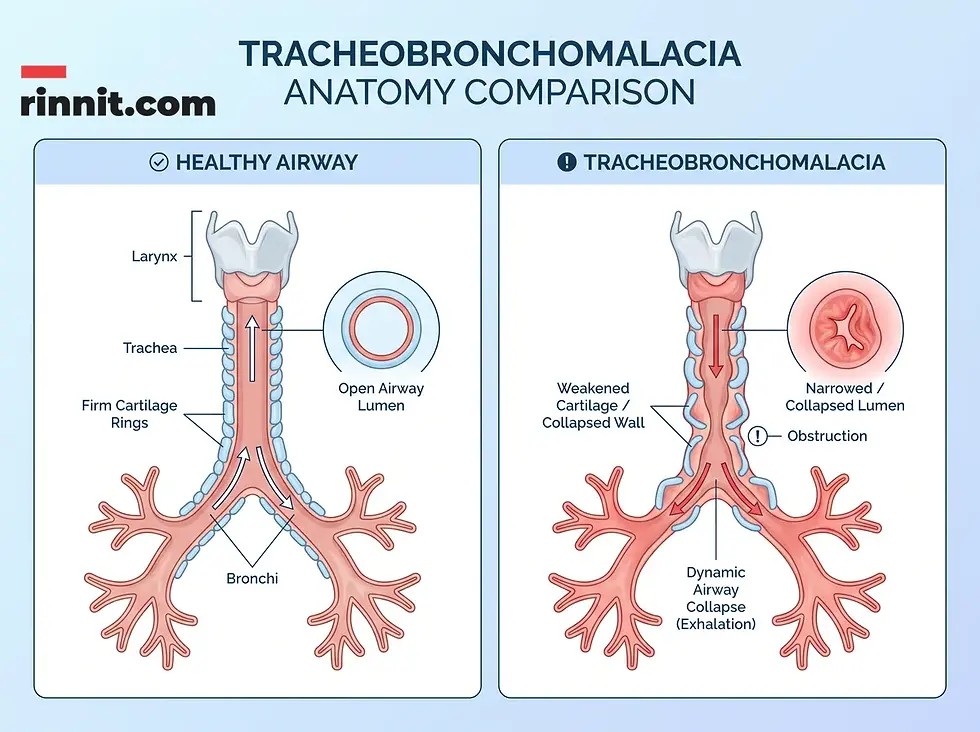
Figure 1: Comparison between a normal, open airway and a collapsed trachea during a TBM event.
Recognizing the Symptoms
The symptoms of TBM often vary depending on the patient's age and the underlying cause of the airway weakness. In infants, signs typically appear between two and four months of age [1]. Adults may experience a gradual progression of symptoms that interfere with daily activities and physical exertion.
Symptom Category | Common Signs in Infants | Common Signs in Adults |
Breathing Sounds | Stridor (high-pitched sound) [1] | Wheezing [1] |
Cough Characteristics | Persistent, chronic cough | Dry, harsh “barking” cough [1] |
Physical Impact | Feeding difficulties [1] | Shortness of breath during activity |
Complications | Frequent respiratory infections [1] | Obstructive sleep apnea (OSA) [1] |
A particularly concerning symptom for adults is the temporary loss of consciousness during intense coughing fits [1]. This occurs because the sudden collapse of the airway can momentarily disrupt oxygen flow and increase internal pressure. Frequent colds and persistent respiratory tract infections are common across all age groups due to the difficulty of clearing mucus from the narrowed passages.
Causes and Risk Factors
The root cause of TBM is the weakening of the cartilage that keeps the airways open. In congenital cases, this may be idiopathic or linked to specific developmental issues. For example, a vascular ring—where the aorta wraps around the trachea [1]—can exert external pressure that weakens the airway walls. Inherited disorders like Ehlers-Danlos syndrome [1] also contribute to cartilage fragility.
Acquired TBM is often the result of chronic inflammation or external pressure. Conditions such as asthma, bronchitis, and COPD [1] are frequent precursors. Obesity and gastroesophageal reflux disease (GERD) are also significant risk factors; nearly half of those treated for severe GERD are found to have some degree of TBM. Environmental toxins, including secondhand smoke and certain toxic gases [1], further increase the risk of developing secondary airway weakness.
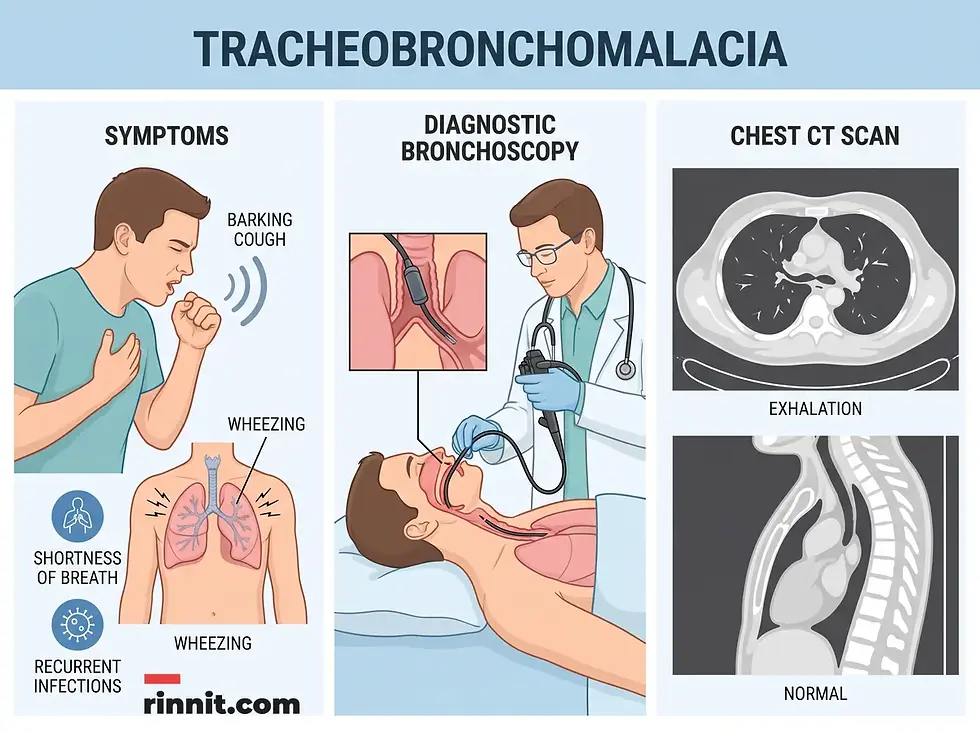
Figure 2: The clinical diagnostic process for TBM, including pulmonary function tests and bronchoscopy.
Diagnostic Procedures
Diagnosing TBM requires a thorough clinical evaluation and specialized testing to visualize the airways in motion. Because the collapse often happens during active breathing or coughing, static imaging alone may not capture the full extent of the problem.
Pulmonary Function Tests: These assess how effectively air moves in and out of the lungs, identifying patterns of obstruction.
Bronchoscopy: This is considered the gold standard for diagnosis. A provider uses a thin camera to observe how much the trachea narrows during deep breaths or coughs.
CT Scans: Advanced imaging helps rule out other potential causes of respiratory symptoms and provides a detailed view of the surrounding anatomy.
Management and Treatment Options
Treatment for TBM is tailored to the severity of the airway collapse and the patient's overall health. For many, the goal is to manage symptoms and prevent complications like infections. In infants, the condition often improves as the cartilage naturally strengthens with age [1], though supportive care is necessary during the interim.
Treatment Type | Description | Primary Goal |
Supportive Devices | CPAP or BiPAP machines [1] | Keep airways open with air pressure |
Mucus Clearance | External percussion vests [1] | Vibrate the chest to loosen mucus |
Medications | Antibiotics or nebulizers [1] | Treat infections and deliver medicine |
Surgical Support | Tracheobronchoplasty | Reinforce airways with mesh [1] |
Surgical intervention is reserved for severe cases where non-invasive methods are insufficient. Procedures like aortopexy involve moving the aorta to relieve pressure on the trachea, while tracheobronchoplasty uses a mesh [1] to provide permanent structural support. Pulmonary rehabilitation is also highly effective, teaching patients specialized techniques to manage their breathing and clear their airways more efficiently.

Figure 3: Overview of TBM management tools, including supportive breathing devices and surgical reinforcement.
Related reading: Several conditions discussed here have their own guides on Rinnit — see Double Aortic Arch: The Most Symptomatic Vascular Ring - Symptoms, Surgery, Recovery and Outlook, Acid Reflux & GERD: Symptoms, Causes, Diagnosis, and Treatment, Chronic Cough: Causes, Symptoms, Diagnosis, and Treatment (2026 Guide), Obstructive Sleep Apnea: Symptoms, Causes, and What Actually Helps and Premature Birth: Complete Guide to Signs, Causes, and Care.
Conclusion
Tracheobronchomalacia is a complex respiratory challenge that requires careful monitoring and a personalized approach to care. Whether congenital or acquired, the condition impacts the fundamental ability to breathe freely. Through a combination of modern diagnostic tools, supportive technology, and advanced surgical techniques, most individuals with TBM can achieve significant symptom relief and maintain a high quality of life.
Call to Action: If you or your child experience a persistent “barking” cough, unexplained wheezing, or significant difficulty breathing during physical activity, consult a healthcare professional for a comprehensive respiratory evaluation. Early identification of airway weakness is the first step toward effective management.
Frequently Asked Questions
1. What exactly is tracheobronchomalacia?
Tracheobronchomalacia is a condition where the windpipe (trachea) and the tubes leading to the lungs (bronchi) collapse during breathing due to weakened cartilage.
2. Can you be born with TBM?
Yes, this is known as primary or congenital TBM. It is often detected in infants around two to four months of age.
3. Is TBM the same as asthma?
No, but they share symptoms like wheezing. TBM is a structural collapse of the airways, while asthma involves inflammation and narrowing.
4. What does a TBM cough sound like?
A TBM cough is often described as a dry, harsh, “barking” cough.
5. Can adults develop TBM?
Yes, acquired TBM can develop due to smoking, obesity, chronic infections, or other underlying health conditions.
6. How common is the congenital form?
Experts estimate that approximately 1 in 2,100 babies are born with the condition.
7. What is a vascular ring?
A vascular ring is a developmental issue where the aorta wraps around the trachea, potentially causing the airway to collapse.
8. How is TBM diagnosed?
The primary method is a bronchoscopy, which allows a provider to see the airway collapse in real-time.
9. Does GERD cause TBM?
Chronic acid reflux is a known risk factor, and nearly half of people treated for severe GERD also have some degree of TBM.
10. What is a percussion vest?
A percussion vest is a device that vibrates the chest to help loosen and clear mucus that gets trapped in collapsed airways.
11. Do all TBM patients need surgery?
No, many cases are managed with supportive devices like CPAP machines or through pulmonary rehabilitation.
12. What is tracheobronchoplasty?
A surgical procedure where a mesh is sewn onto the outside of the airways to provide structural support.
13. Can premature birth increase TBM risk?
Yes, babies born before 37 weeks may have an increased risk if their airway cartilage is not fully formed.
14. Is TBM life-threatening?
While it can cause severe breathing difficulties, it is manageable with proper medical intervention and monitoring.
15. Can TBM go away on its own?
In some infants, the airway cartilage strengthens as they grow, and the condition may improve without major intervention.
References
Cleveland Clinic. Tracheobronchomalacia: Symptoms, Causes & Treatment. Cleveland Clinic. Last reviewed April 1, 2024. https://my.clevelandclinic.org/health/diseases/22061-tracheobronchomalacia
Majid A, Kheir F, Alape D, et al. The prevalence of gastroesophageal reflux in patients with excessive central airway collapse. Chest. 2019;155(3):540-545. PMID 30312588. https://doi.org/10.1016/j.chest.2018.09.021
Mitropoulos A, Song WJ, Almaghlouth F, et al. Detection and diagnosis of large airway collapse: a systematic review. ERJ Open Research. 2021;7(3):00055-2021. PMID 34381840. https://doi.org/10.1183/23120541.00055-2021



Comments