Understanding Retinoschisis: Types, Symptoms, and Management

Updated: 2 days ago
Medically reviewed by Dr. Baraa Alnahhal, MD · Last reviewed: October 17, 2026
Retinoschisis is a condition where the retina divides into two or more layers, potentially impacting central or peripheral vision. [1] While it is a rare disorder with no known cure, management focuses on low vision rehabilitation, refractive correction, and monitoring for complications like retinal detachment.
Quick Answer
Retinoschisis is the splitting of the retina's light-sensing layers, which can affect how the eye sends signals to the brain. It occurs in a congenital form, mostly affecting males, and an acquired form, typically in older adults. [1] Diagnosis involves comprehensive eye imaging. It cannot be cured [1], but vision aids and regular monitoring help maintain quality of life.
What the research says
Most degenerative retinoschisis is benign, and surgery is often unnecessary. A 2022 review of the literature concluded that uncomplicated degenerative retinoschisis (with no breaks in the retinal layers) is almost invariably benign, that retinal detachment linked to outer-layer breaks is fairly common, and that many past interventions were likely unnecessary and may put vision at risk, while progressive symptomatic detachment does need treatment (Ness et al., Survey of Ophthalmology, 2022) [2].
Congenital X-linked retinoschisis stays stable in most eyes, but some progress. In a study of 102 eyes of 51 patients, 53 eyes could be followed without early surgery; over a mean of 7.4 years, 83% (44 eyes) stayed the same type without needing surgery, while 7.5% (4 eyes) developed a combined retinoschisis and retinal detachment that required surgery. The type 3 pattern was a risk factor and may merit closer follow-up (Wood et al., Ophthalmology Retina, 2019) [3].
Myopic traction maculopathy is uncommon overall but more frequent in high myopia. An evidence-based review found that its prevalence is low in the general population but higher among people with high myopia, and that its course tends to be stable, though it can progress and cause worsening vision, while spontaneous improvement also occurs (Cheong et al., Survey of Ophthalmology, 2022) [4].
What is retinoschisis and how does the retina normally work?
Retinoschisis is a medical term derived from 'schisis,' meaning a split or cleft, specifically referring to the division of the retina into multiple layers. [1] The retina is a critical component of the eye responsible for sensing light and sending visual signals to the brain through the optic nerve. [1] When these layers separate, the transmission of light signals is disrupted, which can lead to vision loss in the areas where the split occurs. [1]
The condition primarily affects the light-sensing layer and the cellular layer that communicates with the optic nerve. [1] While these splits can develop in the center of the retina (the macula), they are statistically more likely to occur at the periphery, or the outer edges, of the retinal tissue. [1] Understanding the distinction between retinoschisis and retinal detachment is vital; while detachment involves the retina pulling away from its supporting tissues, retinoschisis is an internal split within the retinal layers themselves. [1]
Feature | Retinoschisis | Retinal Detachment |
Primary Action | Internal splitting of retinal layers | Retina pulls away from supporting tissue |
Typical Location | Often peripheral, can be central | Can affect any part of the retina |
Diagnosis | Requires specialized imaging and testing (OCT, ERG) | The two can be hard to tell apart, even for an ophthalmologist |
Occurring Together | Both can be present at the same time | Possible, especially in the congenital form |
What are the types of retinoschisis?
Retinoschisis is categorized into two distinct types based on when the condition develops and its underlying causes: acquired and congenital. Each type presents with different risk factors and progression patterns.
Acquired (Degenerative) Retinoschisis
Also known as senile retinoschisis, the acquired form typically affects individuals in their 50s, 60s, and 70s. [1] It is estimated to affect approximately 4% of people over the age of 40. [1] Acquired retinoschisis is further divided into 'flat' (typical) and 'bullous' (reticular) subtypes. The bullous subtype is characterized by blistering and carries a higher risk of developing retinal holes or full retinal detachment. [1]
Congenital (Juvenile X-linked) Retinoschisis
This form is a genetic condition present at birth, primarily affecting males. [1] Mothers carry the mutated gene but rarely develop the condition themselves. [1] Juvenile X-linked retinoschisis is rare, occurring in approximately 1 in 5,000 to 1 in 25,000 individuals. [1] It typically affects both eyes [1] and can lead to significant vision challenges early in life.
Myopic Macular Schisis
A specific variant known as myopic macular schisis (or myopic traction maculopathy) occurs at the fovea, the central point of the macula. [1] This type is specifically associated with individuals who have severe myopia, or nearsightedness. [1] [4]
What are the symptoms and warning signs of retinoschisis?
Symptoms of retinoschisis vary significantly depending on the type and severity of the retinal split. Many individuals with the acquired form may remain entirely asymptomatic for years, only discovering the condition during a routine eye examination.
Common signs and symptoms include:
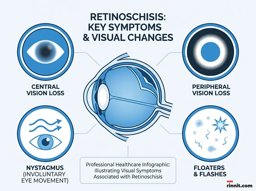
Central and Peripheral Vision Loss: The location of the vision loss depends on where the retinal split occurs. [1]
Congenital Signs: Children may exhibit crossed eyes, uncontrollable side-to-side eye movements (nystagmus), or farsightedness. [1]
Severe Complications: If the condition progresses or leads to retinal detachment, patients may notice floaters, flashes of light, or distorted images. [1]

What causes retinoschisis and what genetic factors are involved?
The causes of retinoschisis differ by type. Congenital retinoschisis is driven by a genetic mutation linked to the X chromosome, explaining why it predominantly affects males. [1] Genetic counseling is often recommended for families with a history of the juvenile form to understand the risks for future generations.
In contrast, the exact cause of acquired retinoschisis remains unknown to scientists. [1] However, age is the most significant risk factor, with the likelihood of development increasing substantially after age 40. [1] While it is often considered a benign condition in its degenerative form [1] [2], regular monitoring is required to prevent progression into more serious retinal issues.
Type | Primary Cause | Key Risk Factor |
Congenital | Genetic mutation (X-linked) | Family history |
Acquired | Unknown | Age (40+) |
Myopic | Retinal traction | Severe nearsightedness |
How is retinoschisis diagnosed?
Diagnosing retinoschisis requires a comprehensive eye exam conducted by an ophthalmologist. [1] Because the condition can mimic retinal detachment, advanced imaging and functional tests are necessary to confirm the diagnosis and assess the extent of the damage.
Standard diagnostic tests include:
Ophthalmoscopy (Fundoscopy): A visual inspection of the retina using a specialized instrument.
Optical Coherence Tomography (OCT): A non-invasive test that uses light to create high-resolution images of the retinal layers.
B-scan Ultrasonography: Uses sound waves to visualize the back of the eye, especially useful if the retina is obscured.
Electroretinography (ERG): Measures the electrical response of the eye to light stimuli to gauge retinal function. [1]
Genetic Testing: Often utilized to confirm juvenile X-linked retinoschisis. [1]
How is retinoschisis managed, and how does vision rehabilitation help?
Retinoschisis has no cure [1], so management focuses on preserving existing vision and addressing complications. The treatment approach is highly individualized based on the patient's symptoms and the type of retinoschisis they possess.
Observation and Refractive Correction
For many patients with acquired retinoschisis, the condition is benign and requires only regular monitoring. [1] [2] Glasses are often prescribed to correct refractive errors such as nearsightedness or farsightedness [1], ensuring the best possible visual clarity.
Addressing Complications
If retinoschisis leads to secondary issues, medical intervention may be required:
Bleeding: Broken blood vessels may be treated with cryoablation (cold therapy) or laser therapy to stop the hemorrhage. [1]
Retinal Detachment: If the split leads to a detachment that is spreading or affecting vision, surgery is needed to re-attach the retinal tissue; small, stable detachments may only be watched. [1]
Low Vision Support: Patients with significant vision loss benefit from magnifiers, adaptive technology, and mobility training.
Emerging Research
Scientists are actively researching potential new treatments, particularly for the congenital form. These include gene therapy, stem-cell therapy, and the use of specific medications like dorzolamide to manage the condition. [1]

Conclusion and Next Steps
Retinoschisis is a complex retinal disorder that requires professional diagnosis and long-term management. While the lack of a cure can be challenging, most cases can be effectively managed through regular eye examinations, vision aids, and prompt treatment of complications. Education and adaptive technologies play a crucial role in helping those with significant vision loss maintain their independence and quality of life.
Next step: If you or your child are experiencing changes in vision, such as new floaters, flashes, or peripheral vision loss, schedule a comprehensive eye exam immediately. Early detection and regular monitoring are the most effective ways to protect your long-term retinal health.
Frequently Asked Questions
What is the main difference between retinoschisis and retinal detachment?
Retinoschisis is an internal split within the layers of the retina itself, whereas retinal detachment occurs when the entire retina pulls away from the underlying supporting tissues.
Can retinoschisis be cured?
No, there is currently no cure for retinoschisis. Management focuses on correcting vision with glasses, using low vision aids, and monitoring for complications.
Who is most at risk for congenital retinoschisis?
Congenital (juvenile X-linked) retinoschisis primarily affects males, as it is a genetic condition carried by the mother on the X chromosome.
Is acquired retinoschisis a sign of aging?
Yes, acquired retinoschisis is often called senile retinoschisis because it most commonly affects people in their 50s, 60s, and 70s.
What is bullous retinoschisis?
Bullous retinoschisis is a subtype of the acquired form characterized by blistering of the retinal layers. It carries a higher risk of progressing to retinal detachment.
How common is the juvenile form of the disease?
Juvenile X-linked retinoschisis is relatively rare, occurring in approximately 1 in 5,000 to 1 in 25,000 individuals.
What tests are used to diagnose retinoschisis?
Diagnosis typically involves ophthalmoscopy, OCT imaging, B-scan ultrasound, and electroretinography (ERG) to assess retinal function.
Can glasses help with retinoschisis?
While glasses cannot fix the retinal split, they are often used to correct refractive errors like nearsightedness or farsightedness that may accompany the condition.
What are 'floaters and flashers' in the context of this disease?
Floaters and flashers are often signs that retinoschisis has become severe or that a retinal detachment is occurring, requiring immediate medical attention.
Are there new treatments on the horizon?
Researchers are currently investigating gene therapy, stem-cell therapy, and certain medications as potential future treatments for the congenital form.
Related Reading
Retinal Detachment: Symptoms, Causes, Diagnosis & Treatment — Complete Guide explains retinal detachment, the main complication of retinoschisis.
Retinal Tear: Symptoms, Causes, and Treatment Options covers retinal tears and the warning signs that need urgent care.
Optical Coherence Tomography (OCT) Eye Test: What It Shows and What to Expect describes the eye scan used to see splitting in the retina.
Retina: Function, Anatomy, Symptoms, Tests and Care explains how the retina works and common tests.
References
Cleveland Clinic. Retinoschisis. Cleveland Clinic. October 18, 2022. https://my.clevelandclinic.org/health/diseases/24310-retinoschisis
Ness S, Subramanian ML, Chen X, et al. Diagnosis and management of degenerative retinoschisis and related complications. Survey of Ophthalmology. 2022;67(4):892-907. PMID 34896193. https://doi.org/10.1016/j.survophthal.2021.12.004
Wood EH, Lertjirachai I, Ghiam BK, et al. The natural history of congenital X-linked retinoschisis and conversion between phenotypes over time. Ophthalmology Retina. 2019;3(1):77-82. PMID 30935660. https://doi.org/10.1016/j.oret.2018.08.006
Cheong KX, Xu L, Ohno-Matsui K, et al. An evidence-based review of the epidemiology of myopic traction maculopathy. Survey of Ophthalmology. 2022;67(6):1603-1630. PMID 35367479. https://doi.org/10.1016/j.survophthal.2022.03.007
Medical disclaimer
The information provided in this article is for educational purposes only and should not be used as a substitute for professional medical advice, diagnosis, or treatment. Always seek the advice of your physician or other qualified health provider with any questions you may have regarding a medical condition.



Comments