Understanding Melanonychia: Causes, Symptoms, and Diagnosis

Updated: 3 days ago
Medically reviewed by Dr. Baraa Alnahhal, MD · Last reviewed: October 2026
Melanonychia is a brown or black discoloration of a fingernail or toenail caused by melanin deposits in the nail plate. It is often harmless, but it can sometimes signal an underlying health problem or skin cancer[1].
Quick Answer: What is melanonychia?
Melanonychia is a darkened area, usually brown or black, in a fingernail or toenail, caused when pigment cells deposit melanin into the nail plate. It is common and often harmless, particularly in people with medium to dark skin tones, but it can occasionally signal subungual melanoma. A new or changing streak should be evaluated, and treatment depends on the cause. [1]
What the research says
A single dark band on one adult nail usually calls for a biopsy. A clinical review of melanonychia diagnosis states that dermoscopy is used routinely, and that a biopsy with histopathologic examination is the gold standard when melanocytic proliferation is suspected. A biopsy is recommended when longitudinal melanonychia occurs in an adult, affects a single digit, and has no local or systemic cause that explains it (Alessandrini et al., Dermatol Clin, 2021 [2]).
The ABCDEF checklist cannot replace a biopsy for a worrying band. In a single-center study of 84 biopsied cases of longitudinal melanonychia (8 subungual melanomas and 76 benign), melanomas more often had a wider band and a greater band-width percentage, but the number of ABCDEF criteria met did not differ between the groups. The authors advise biopsy for any concerning band, especially one with a width percentage above 40% (Ko et al., J Am Acad Dermatol, 2019 [3]).
Skin tone changes how benign bands look and how often they are biopsied. In a 10-year retrospective cohort of 248 benign longitudinal melanonychia cases, 47 patients (18.95%) had a nail matrix biopsy. Darker-skinned patients had wider, darker bands, more band changes, and more biopsies than lighter-skinned patients (Lee et al., J Am Acad Dermatol, 2022 [4]).
What Is Melanonychia?
Melanonychia refers to a darkened area in the nail plate of a fingernail or toenail. It occurs when melanin, the substance that gives hair, eyes, and skin their color, is deposited into the nail plate. The nail plate itself is made of keratin, which has no color. Cells that contain melanin are present in the nails but are usually inactive, and certain situations and conditions can activate them[1].
The condition is common and often harmless. It is particularly frequent in people with medium to dark skin tones, where it is often a benign genetic trait. However, because melanonychia can also be a sign of subungual melanoma, a serious form of skin cancer, any new or changing nail discoloration should be evaluated by a healthcare provider[1].
What are the types and symptoms of melanonychia?
The main symptom is an area of dark color in a fingernail or toenail. It may be brown, gray, or black, and it can affect one nail or several. Most often it appears as a vertical band that can look like paint or marker residue, but it does not wash off[1].
Type of melanonychia | Visual presentation |
Longitudinal | A vertical darkened line running from the cuticle to the tip of the nail. This is the most common type. |
Transverse | A horizontal darkened band across the width of the nail plate. |
Total (diffuse) | Darkening that covers the entire nail plate. |
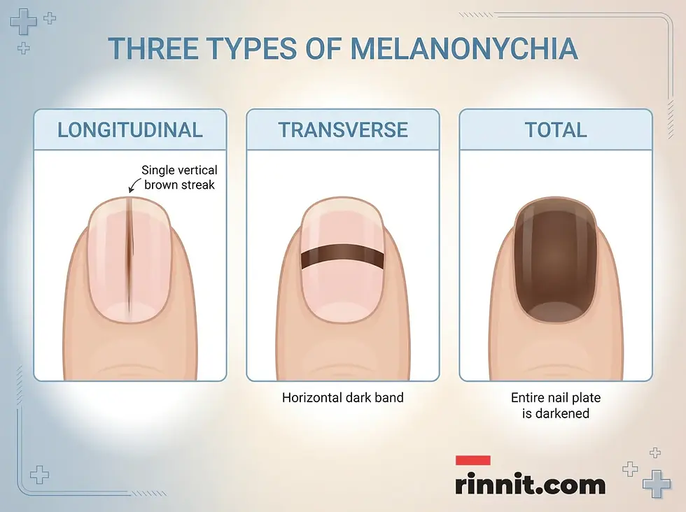
Melanonychia can appear as a vertical streak, a horizontal band, or a fully darkened nail.
Other symptoms depend on the cause. Trauma, infection, and inflammatory conditions can damage the nail so that it splits or becomes painful. If a streak is cancerous, the pigment may spread beyond the nail onto the surrounding skin, a sign known as Hutchinson's sign[1].
What commonly causes melanonychia, and what are the risk factors?
The most common causes are noncancerous. They include a bruised nail, a mole (nevus) in the nail, repeated nail trauma such as nail biting, picking, or friction from tight shoes, lentigos (age spots) in the nail, and genetics. People with medium to dark skin tones, who have more melanin, often develop melanonychia[1].

Trauma, benign moles, and genetics are common noncancerous causes.
Other causes include cancerous tumors such as subungual melanoma, and a range of health conditions and exposures[1]:
Infections: fungal or bacterial nail infections, warts, and HIV.
Connective tissue and inflammatory conditions: scleroderma, lupus, psoriasis, lichen planus, and amyloidosis.
Hormone conditions: Addison's disease, Cushing's syndrome, and an overactive thyroid.
Medications and procedures: chemotherapy, antiretroviral therapy (ART), antimalarials, phototherapy, and X-ray exposure.
Nutritional deficiencies: such as vitamin B12 deficiency.
Pregnancy.
Who Is Most Affected?
Melanonychia is most common in people with medium to dark skin tones. Among people of Caribbean descent, nail discoloration by age 50 is common, and nearly 20% of people of Japanese descent have benign melanonychia[1].
How is melanonychia diagnosed?
Melanonychia is diagnosed with a physical exam and a review of your symptoms and medical history, using the ABCDEF method to judge cancer risk. They will ask questions based on the ABCDEF method, which helps judge the risk of cancer[1][3]:
A (Age): your age and demographic risk factors.
B (Band): the width and borders of the pigmented band.
C (Changes): changes in the size or shape of the line, and how long it took to appear.
D (Digits): which fingers or toes are involved.
E (Extension): whether the pigment has spread to the surrounding skin.
F (Family history): a family history of melanoma or atypical moles.
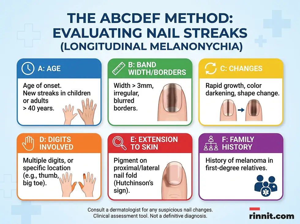
The ABCDEF method helps providers assess nail streaks. It is a clinical tool, not a definitive diagnosis.
To find out whether a nail change is noncancerous or cancerous, your provider may look at a nail clipping under a microscope, examine the nail with a handheld microscope (dermoscopy), take a biopsy of a small sample of nail and skin, or order imaging tests to look below the skin for signs of cancer[1][2].
How is melanonychia managed and treated?
Treatment depends on the cause. If the streak is benign and only cosmetic, you may not need any treatment. If an underlying condition is responsible, that condition is treated, for example with medication for an infection. If the diagnosis is subungual melanoma, you will need cancer therapies, which may include surgery[1].
In most cases melanonychia is not serious. If it is caused by your race or ethnicity, you will likely have it for life. If a noncancerous underlying condition is the cause, it may fade after a few months of treatment. Melanoma in the nail generally has a poorer outlook than melanoma on other skin, so early diagnosis and treatment make a big difference[1].
What is the bottom line on melanonychia?
Melanonychia is a common nail condition that is usually harmless, but it needs careful attention to rule out serious causes. Whether it appears as a thin vertical line or a broad band of color, the key is an accurate diagnosis. If you notice a new or changing dark streak on a fingernail or toenail, do not ignore it. Schedule an appointment with a healthcare provider or dermatologist for an evaluation[1].
Frequently Asked Questions
What exactly is melanonychia?
Melanonychia is a brown or black discoloration of a fingernail or toenail caused by a deposit of melanin in the nail plate.
Is a dark line on the nail always cancer?
No. Most cases are harmless and can be caused by genetics, trauma, or a benign mole, although a dark streak can sometimes be a sign of melanoma.
Can nail biting cause dark streaks?
Yes. Repeated nail trauma, including biting or picking, can activate melanin-producing cells and lead to nail discoloration.
Who is most likely to develop melanonychia?
Melanonychia is most common in people with medium to dark skin tones, including people of Caribbean and Japanese descent.
Does melanonychia wash off?
No. The pigment is embedded in the nail plate, so it cannot be washed or scrubbed away like residue on the surface.
What is the ABCDEF method for diagnosis?
The ABCDEF method is a clinical tool that assesses a nail streak by Age, Band characteristics, Changes, Digits involved, Extension of pigment to the surrounding skin, and Family history of melanoma or atypical moles.
Can pregnancy cause nail discoloration?
Yes. Pregnancy is one of the known causes of melanonychia.
How is subungual melanoma different?
Subungual melanoma is a type of skin cancer that develops in the nail area. It is one of the possible causes of melanonychia and needs prompt diagnosis and treatment.
Is treatment always necessary for melanonychia?
No. If the streak is benign and only cosmetic, treatment is usually not needed. An underlying infection, condition, or cancer does need treatment.
Can vitamin deficiencies affect nail color?
Yes. A vitamin B12 deficiency is a known nutritional cause of melanonychia.
Related Reading
Melanoma: A Comprehensive Guide to Early Detection and Treatment explains how to spot melanoma early and how it is treated.
Skin Biopsy: Types, What to Expect, Results and Aftercare describes how a tissue sample is taken to check for cancer.
Nail Trephination: Procedures for Draining Subungual Hematomas explains how trapped blood under a nail is drained.
Onychomycosis: Nail Fungus Symptoms, Diagnosis and Treatment covers nail fungus, a common cause of discolored nails.
References
Cleveland Clinic. Melanonychia. Cleveland Clinic. November 10, 2025. https://my.clevelandclinic.org/health/diseases/melanonychia
Alessandrini A, Dika E, Starace M, Chessa MA, Piraccini BM. Diagnosis of melanonychia. Dermatol Clin. 2021;39(2):255-267. PMID 33745638. https://doi.org/10.1016/j.det.2020.12.004
Ko D, Oromendia C, Scher R, Lipner SR. Retrospective single-center study evaluating clinical and dermoscopic features of longitudinal melanonychia, ABCDEF criteria, and risk of malignancy. J Am Acad Dermatol. 2019;80(5):1272-1283. PMID 30765143. https://doi.org/10.1016/j.jaad.2018.08.033
Lee DK, Chang MJ, Desai AD, Lipner SR. Clinical and dermoscopic findings of benign longitudinal melanonychia due to melanocytic activation differ by skin type and predict likelihood of nail matrix biopsy. J Am Acad Dermatol. 2022;87(4):792-799. PMID 35752275. https://doi.org/10.1016/j.jaad.2022.06.1165
Medical disclaimer
This article is for informational purposes only and is not a substitute for professional medical advice, diagnosis, or treatment. The clinical information in this guide reflects source material last reviewed in November 2025.



Comments