Thoracostomy (Chest Tube Insertion): Purpose, Procedure, Risks and Recovery

Updated: 6 days ago
Written and medically reviewed by Dr. Baraa Alnahhal, MD · Last reviewed: October 2026 · How we review
Quick Answer: What is a thoracostomy?
A thoracostomy is a procedure in which a provider makes a small cut between two ribs and places a chest tube to drain air or fluid from the pleural space around the lung. It treats collapsed lung, pleural effusion, empyema, hemothorax and chylothorax, uses local numbing medicine, and usually takes about 30 minutes. [1]
What the research says
About 1 in 5 chest tubes placed after an injury leads to a complication. A review of 29 studies covering 4,981 chest tubes in trauma patients found a median complication rate of 19%. Problems with tube position made up 53.1% of complications, and the overall rate stayed stable across three decades. (Hernandez et al., J Trauma Acute Care Surg, 2018) [2]
Smaller chest tubes drained blood after chest injury about as well as larger ones. Across 9 studies and 1,847 patients, the failure rate was 17.8% with small tubes (14 Fr or less) and 21.5% with large tubes (20 Fr or more), a difference that was not significant. Small tubes stayed in for fewer days on average (4.3 vs 6.2). (Lyons et al., J Trauma Acute Care Surg, 2024) [3]
Not every collapsed lung needs a chest tube. A review of 22 studies on first-time spontaneous collapsed lung found that chest tubes cleared the collapse more often without another procedure, but careful observation meant shorter hospital stays, the same two-year recurrence rate, and the best value in 98.2% of cost simulations. (Eamer et al., Chest, 2023) [4]
Key takeaways
A thoracostomy is the procedure; the chest tube is the device it places. [1]
The tube drains air, blood, pus, lymph or other fluid from the space between the lung and the chest wall. [1]
A needle thoracostomy is an emergency step for a tension pneumothorax and is usually followed by a tube thoracostomy. [1]
The procedure is minimally invasive, uses local anesthesia, and usually takes about 30 minutes. [1]
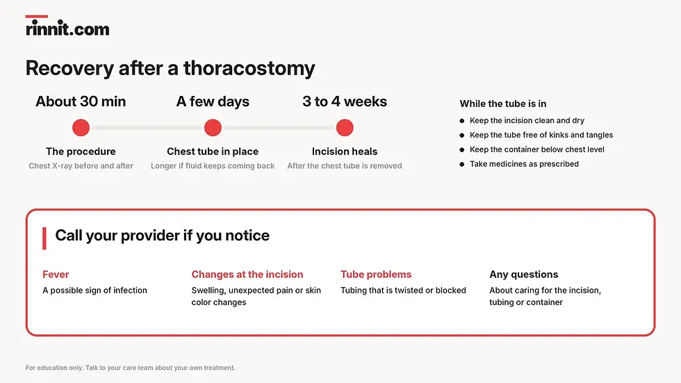
Most chest tubes stay in for a few days; the incision may take three to four weeks to heal after removal. [1]
Fever, swelling, unexpected pain or skin color changes at the incision, or a twisted or blocked tube, need a call to your provider. [1]
Thoracostomy at a glance
Feature | What to know |
What it is | A small incision in the chest to insert a chest tube [1] |
Purpose | Drains air or fluid from the pleural space around the lung [1] |
Used for | Emergencies and long-term (chronic) conditions [1] |
Anesthesia | Local anesthetic (such as lidocaine); no general anesthesia needed [1] |
Imaging | Chest X-ray or other imaging before, and a chest X-ray after [1] |
Procedure time | About 30 minutes [1] |
How long the tube stays | Usually a few days; longer if fluid keeps coming back [1] |
Incision healing | Three to four weeks after the tube is removed [1] |

What is a thoracostomy and how does a chest tube work?
A thoracostomy is a procedure in which a surgeon makes an incision and inserts a tube into the chest to drain air or fluid from the pleural space. [1]
The pleural space sits between two thin membranes called the pleurae. One lines the inside of the chest wall and the other covers the lung. [1] When air, blood, pus or fluid collects in this space, it can press on the lung. The chest tube gives that build-up a way out, and the tube may connect to a drainage container. [1]
Providers use thoracostomy in two very different settings: sudden emergencies and ongoing (chronic) conditions where fluid keeps returning. [1] For a deeper look at the structures involved, see our guides to the chest (thorax) and the rib cage.
What conditions does a thoracostomy treat?
A thoracostomy treats conditions in which air or fluid builds up around the lungs, including collapsed lung, pleural effusion, empyema, hemothorax, chylothorax and lung infections. [1]
What is the difference between a needle thoracostomy and a tube thoracostomy?
A needle thoracostomy is a fast emergency procedure for a tension pneumothorax, while a tube thoracostomy places a chest tube that keeps draining over time. [1]
In a tension pneumothorax, air can get into the space around the lung but cannot get out. [1] A needle placed between two ribs in the upper chest releases that trapped air until a chest tube can be inserted. [1]
Feature | Needle thoracostomy | Tube thoracostomy |
Main use | Emergency treatment of tension pneumothorax [1] | Draining air or fluid in emergencies or chronic conditions [1] |
Insertion site | Upper chest, between two ribs [1] | Side of the chest, between two ribs [1] |
What is inserted | A needle [1] | A tube, often attached to a drainage container [1] |
What comes next | Usually followed by a tube thoracostomy [1] | Care of the tube and dressing until removal [1] |
How do you prepare for a thoracostomy?
Preparation for a thoracostomy depends on whether it is planned, because an emergency thoracostomy happens without time to prepare. [1]
For a planned procedure, your provider may ask you to: [1]
Stop certain medications before the procedure.
Change your medication schedule in the days leading up to it.
Avoid eating or drinking for a set time beforehand.
Wear comfortable clothes on the day.
Tell your provider: [1]
Every medication you take, including vitamins and supplements.
If you are pregnant or think you could be.
If you are allergic to any medications.
What happens during a tube thoracostomy?
A tube thoracostomy follows a set sequence: imaging, positioning, cleaning, numbing, a small cut between two ribs, tube insertion and a sterile dressing. [1]
Before the procedure, you will have a chest X-ray or another imaging test so the provider knows exactly where to place the tube. [1] Then the provider will: [1]
Position you with your head raised and one arm lifted beside your head.
Clean and sterilize an area on the side of your chest.
Numb the area with a local anesthetic such as lidocaine, then wait a few minutes to make sure you are numb.
Make a small incision on the side of your chest, between two ribs.
Attach a clamp (hemostat) that looks like scissors or pliers to the opening — this step is not done in every thoracostomy.
Insert the tube through the incision into your chest; it may connect to a drainage container.
Apply a sterile dressing around the incision.
For a needle thoracostomy, the provider cleans and sterilizes an area on the upper chest and inserts a needle between two ribs. [1]
The whole procedure usually takes about 30 minutes. [1]

What happens after a thoracostomy?
After a thoracostomy, you will have another chest X-ray and receive instructions on caring for yourself and the chest tube. [1]
Your care instructions may include how to: [1]
Keep the skin at the incision site clean and dry.
Look after the tube and drainage container, making sure the tube does not kink or tangle.
Keep the drainage container below chest level.
Take any medications your provider prescribes.
What are the benefits and risks of a thoracostomy?
The main benefits of a thoracostomy are that it is minimally invasive and avoids general anesthesia, while its main risks include tube misplacement, infection, bleeding and tissue damage. [1]
Benefits | Risks and complications |
Minimally invasive [1] | Tube misplacement or dislodging [1] |
Useful in emergencies and for chronic conditions [1] | Infection [1] |
No general anesthesia needed [1] | Subcutaneous emphysema (air trapped under the skin) [1] |
Can make you more comfortable when fluid around the lungs keeps returning [1] | Re-expansion pulmonary edema (fluid in the lung after it reinflates) [1] |
— | Bleeding [1] |
— | Nerve or tissue damage [1] |
Every procedure carries some risk, and your care team takes precautions to keep these risks as low as possible. [1] Research in injured patients puts the overall complication rate at about 19%, with tube position problems being the most common type. [2]
How long does a chest tube stay in after a thoracostomy?
A chest tube placed during a thoracostomy usually stays in only until all the fluid drains, which typically takes a few days. [1]
If fluid keeps coming back because of a chronic condition, the tube may stay in much longer. [1] Your provider will tell you what to expect in your situation. [1]
How long does recovery from a thoracostomy take?
Recovery from a thoracostomy mainly involves healing of the incision, which may take three to four weeks after the chest tube is removed. [1]
Your provider will explain how to care for the incision site and whether you need to limit any activities while it heals. [1]
When should you call a provider after a thoracostomy?
You should call your provider after a thoracostomy if you notice signs of infection, if the tubing becomes twisted or blocked, or if you are unsure how to care for the tube or incision. [1]
Contact your provider if you have: [1]
Signs of infection: fever, swelling or unexpected pain at the incision site, or changes in skin color.
Tube problems: tubing that is twisted or blocked. If the tube falls out or comes apart, or you suddenly have chest pain or trouble breathing, call 911 or your local emergency number.
Questions: anything you are unsure about in caring for the incision, the tubing or the drainage container.
How is a thoracostomy different from thoracentesis and thoracotomy?
A thoracostomy places a chest tube that drains over several days, while thoracentesis drains fluid or air in a single procedure and thoracotomy is open surgery on the chest. [1]
Procedure | What it does |
Thoracentesis | Drains fluid or air from the chest [1] |
Thoracostomy | A minimally invasive procedure that inserts a chest tube to drain fluid or air over a few days [1] |
Thoracotomy | A more invasive surgery that lets a surgeon see inside the chest cavity to diagnose or treat illness; a chest tube may be placed afterward [1] |
Related reading: pleurisy and chest pain.
Conclusion
A thoracostomy is a quick, minimally invasive procedure that places a chest tube to drain air, blood, pus, lymph or fluid from around the lung. [1] It can be life-saving in an emergency and can bring lasting comfort when fluid keeps returning because of a chronic condition. [1] Most tubes stay in for a few days, and the incision usually heals within three to four weeks of removal. [1]
If you or someone you care for is scheduled for a chest tube, write down your questions and bring them to your care team — they can explain what to expect and address any concerns. [1]
Keep learning: explore our clear, medically reviewed guides to collapsed lung, hemothorax and pneumonia, and share this article with anyone preparing for a chest tube.
FAQ
Is a thoracostomy the same as a chest tube?
A thoracostomy is the procedure, and the chest tube is the device it places. The chest tube then drains fluid or air that builds up in the chest because of certain medical conditions. [1]
Is a thoracostomy painful?
A thoracostomy is done with a local anesthetic, such as lidocaine, that numbs the side of the chest before any incision is made. General anesthesia is not needed, and the provider waits a few minutes to make sure the area is numb. [1]
How long does a thoracostomy take?
A thoracostomy usually takes about 30 minutes. A chest X-ray is done before the procedure to guide tube placement and again afterward. [1]
How long does a chest tube stay in?
A chest tube usually stays in for a few days, until all the fluid drains. A chest tube may stay in much longer if fluid keeps returning because of a chronic condition. [1]
How long does a thoracostomy incision take to heal?
A thoracostomy incision may take three to four weeks to heal completely after the chest tube is removed. Your provider will explain how to care for the site and whether to limit activities. [1]
What are the risks of a thoracostomy?
The risks of a thoracostomy include tube misplacement or dislodging, infection, air under the skin, fluid in the lung after it reinflates, bleeding, and nerve or tissue damage. Problems with tube position are the most common complication reported after chest injury. [1][2]
When is a needle thoracostomy used?
A needle thoracostomy is used in emergencies to treat a tension pneumothorax, when air enters the space around the lung but cannot escape. A needle thoracostomy is usually followed by a tube thoracostomy. [1]
When should I call my provider after a thoracostomy?
Call your provider after a thoracostomy if you have fever, swelling, unexpected pain or skin color changes at the incision, or if the tubing is twisted or blocked. Also call with any questions about caring for the tube, incision or drainage container. [1]
Are you awake during chest tube insertion?
Often, yes. The provider numbs the skin and chest wall, and you may get medicine to help you relax. A chest tube may also be placed during surgery while you are under general anesthesia.
How is a chest tube removed?
The provider removes the stitches and pulls the tube out quickly, often while you hold your breath or breathe out. A dressing covers the site, and you may have a chest X-ray afterward.
Related Reading
Learn more in our guide to Bullectomy.
References
Cleveland Clinic. Thoracostomy: Purpose, Procedure & Risks. Cleveland Clinic. Last updated May 31, 2023. https://my.clevelandclinic.org/health/treatments/25035-thoracostomy
Hernandez MC, El Khatib M, Prokop L, et al. Complications in tube thoracostomy: Systematic review and meta-analysis. J Trauma Acute Care Surg. 2018;85(2):410-416. PMID 29443856. https://doi.org/10.1097/TA.0000000000001840
Lyons NB, Abdelhamid MO, Collie BL, et al. Small versus large-bore thoracostomy for traumatic hemothorax: A systematic review and meta-analysis. J Trauma Acute Care Surg. 2024;97(4):631-638. PMID 39213292. https://doi.org/10.1097/TA.0000000000004412
Eamer G, Povolo CA, Petropoulos JA, et al. Observation, Aspiration, or Tube Thoracostomy for Primary Spontaneous Pneumothorax: A Systematic Review, Meta-Analysis, and Cost-Utility Analysis. Chest. 2023;164(4):1007-1018. PMID 37209773. https://doi.org/10.1016/j.chest.2023.05.017
Further reading (professional): Dezube R. How To Do Tube and Catheter Thoracostomy. Merck Manual Professional Version. Last updated September 2026. https://www.merckmanuals.com/professional/pulmonary-disorders/how-to-do-pulmonary-procedures/how-to-do-tube-and-catheter-thoracostomy
Medical disclaimer
This article is for general educational purposes only and does not replace professional medical advice, diagnosis or treatment. Always ask your doctor or another qualified healthcare provider about any questions you have regarding a medical condition or procedure. If you have sudden chest pain or trouble breathing, seek emergency care immediately.


Comments