Pericardiocentesis: Procedure, Risks, and Recovery

Updated: 3 days ago
Medically reviewed by Dr. Baraa Alnahhal, MD · Last reviewed: October 2026
Pericardiocentesis is a procedure that drains extra fluid from the pericardium, the protective pouch around the heart. It can be life-saving when so much fluid builds up that the heart no longer has room to beat properly, a condition called cardiac tamponade. A provider uses a needle and imaging guidance to reach the fluid, relieve the pressure, and, when needed, collect a sample to find the cause [1].
Quick Answer
Pericardiocentesis is a procedure that removes excess fluid from around the heart using a needle placed into the pericardium with imaging guidance, usually ultrasound. It takes about 10 to 20 minutes and can be done as emergency treatment or as a diagnostic test. Success rates are higher than 90%, and complication risk is lowest when imaging is used. [1]
What the research says
Imaging-guided drainage is highly successful. In a review of 66 patients at one hospital (62.1% with active malignancy), pericardiocentesis succeeded in 65 (98.5%); 2 patients (3%) developed an access-site infection and 1 (1.5%) became hemodynamically unstable. About 70% of procedures used echocardiography and 27% used fluoroscopy (Baqi & Ahmed, Cureus, 2020).
A sitting-position apical approach worked well in cancer patients. In 53 consecutive patients with moderate or large malignant effusions, high-frequency ultrasound-guided drainage from the apical approach in the sitting position succeeded in 100%, with no major complications and minor complications in 8%; 11% had a recurrent effusion within 90 days. This was a single-center series (Zhang et al., Clin Cardiol, 2021).
Effusions can come back in people with cancer. Among 418 cancer patients who had a drain placed for a large effusion, 65 (16%) needed further drainage for a recurrence, and the cumulative incidence was 15.0% at 12 months (Ahmed et al., Front Cardiovasc Med, 2022).
Research summaries are based on article abstracts retrieved from PubMed.
Why is pericardiocentesis needed?
A small amount of fluid in the pericardium is normal. Too much fluid, called pericardial effusion, can lead to cardiac tamponade, a medical emergency that can stop the heart and be deadly within minutes to hours [1]. In emergencies, pericardiocentesis treats tamponade or severe effusions that are about to cause it. In non-emergencies, it is often used as a diagnostic test to find out why fluid is building up [1].
The procedure is relatively common: providers in the United States perform more than 29,000 of them each year [1].

What are the common causes of pericardial effusion needing pericardiocentesis?
Cause | Description |
Advanced cancer | Cancer that has spread to the heart or pericardium, and heart cancer. |
Heart attack | Especially when the heart wall ruptures. |
Infections and inflammation | Bacterial or viral infections of the pericardium, including after COVID-19 or with HIV, and tuberculosis (more common in developing countries). |
Chronic immune diseases | Lupus, rheumatoid arthritis, and scleroderma. |
Organ failure | Heart failure or kidney failure. |
Underactive thyroid | Hypothyroidism. |
Medical treatments | Certain drugs or radiation therapy. |
Unknown cause | Some effusions are idiopathic. |
How do you prepare for pericardiocentesis?
Preparing for pericardiocentesis depends on whether the procedure is an emergency or planned in advance. For a planned procedure you will need to fast starting eight hours beforehand, though clear liquids are allowed until two hours before. Your care team will [1]:
Start an intravenous (IV) line for medications or fluids.
Remove hair at the needle site and clean the skin with antiseptic, often preparing several areas.
Set up monitoring of blood pressure, breathing, heart rate, and blood oxygen, sometimes with ECG electrodes that help guide needle placement.
Give extra oxygen if your blood oxygen is low or might drop.
What happens during a pericardiocentesis procedure?
Pericardiocentesis is performed by a team of doctors, nurses and imaging technicians, who use imaging to guide a needle into the pericardium and draw out fluid. Ultrasound (echocardiogram) is the most common guidance, and fluoroscopy is another option. Imaging helps place the needle exactly where it needs to go. Doing the procedure without imaging is extremely rare, reserved for extreme emergencies, and carries a higher risk of complications [1].
Unless the heart is about to stop, a local anesthetic numbs the skin first, and a small skin cut may be made to ease needle insertion. There are three entry points [1]:
Entry Approach | Location and Access |
Substernal | Under the breastbone; the most common route, usually giving easy, direct access to the pericardium. |
Parasternal | Just off-center at the front of the chest, going between the ribs from the front. |
Apical | Between the ribs from the side of the body, aiming for the pointed end (apex) of the heart. |
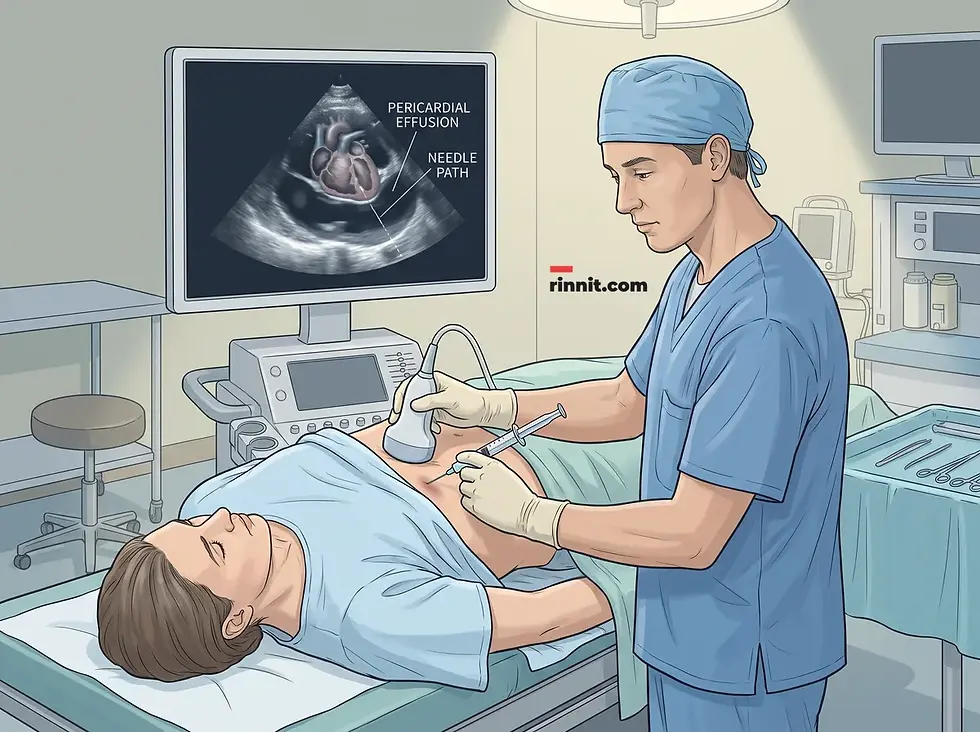
Once the needle tip is in the pericardium, fluid is drawn out. The procedure takes about 10 to 20 minutes. If there is a lot of fluid, a catheter may be placed to drain it more slowly and can be left in for a day or two. Afterward the site is bandaged [1].
What happens after pericardiocentesis?
Your provider may test the fluid to find the cause, repeat the ultrasound to confirm the fluid is gone (daily if a drain is in place), and remove the drain once the fluid has cleared [1].
What are the benefits, success rate and risks of pericardiocentesis?
Besides being life-saving when fluid is squeezing the heart, pericardiocentesis can reveal the cause of the fluid, can be done in a hospital room, operating room, or cardiac catheterization lab, and is faster and less invasive than surgery. Studies report success rates higher than 90%, with some as high as 100%. Fluid can build up again, and if it does, a surgeon may discuss a more permanent procedure called a pericardial window [1].
Complications happen in about 5% to 40% of cases, and the risk is lowest when imaging guides the needle. Because the needle passes close to vital organs, possible injuries include [1]:
Heart: bleeding, a heart attack, or a disrupted heart rhythm, which can be severe and may need emergency surgery.
Lungs: a puncture that causes the lung to collapse.
Other structures: the diaphragm, pericardium (fluid leak or air in the pericardium), liver, stomach, or major blood vessels near the heart.
Infection: which can spread and lead to sepsis, a life-threatening emergency.
Pain is usually limited: the numbing medicine may sting, and you may feel pressure when the needle enters the pericardial sac [1].
What are recovery and the outlook after pericardiocentesis?
Most people start to feel better quickly, often right after the fluid is drained. You will need to rest for 12 to 24 hours while providers watch your condition, and the overall recovery time depends on how severe the problem was, what caused it, and any other treatments you receive. Follow-up visits check for complications or a need for more treatment, and some people need more than one procedure because effusions can recur, especially with certain cancers or infections such as tuberculosis [1].

When to Go to the Hospital
After the procedure, seek care right away for [1]:
Signs of cardiac tamponade: chest pain, trouble breathing or breathing unusually fast, fainting, dizziness or lightheadedness, or heart palpitations or a rapid heartbeat.
Signs of infection or sepsis: swelling, redness, or unusual warmth around the needle site, fever or chills, or confusion or disorientation.
Conclusion
Pericardiocentesis is a proven, often life-saving way to relieve pressure on the heart and to find out why fluid collected there. With imaging guidance, outcomes are usually good, and the potential to save lives almost always outweighs the risks. Ask your provider about your specific situation [1].
Frequently Asked Questions
What is the main purpose of pericardiocentesis?
To drain excess fluid from the pericardium, relieving pressure on the heart and preventing or treating cardiac tamponade, and sometimes to find the cause of the fluid [1].
Is pericardiocentesis an emergency procedure?
Pericardiocentesis can be either. In emergencies it treats tamponade or severe effusions about to cause it; in non-emergencies it is often used as a diagnostic test [1].
How long does the procedure take?
About 10 to 20 minutes, although preparation and recovery take longer [1].
What kind of anesthesia is used?
Usually a local anesthetic that numbs the needle site. In the most extreme emergencies it may be skipped [1].
Why is imaging guidance used?
Ultrasound or fluoroscopy lets the provider place the needle exactly where it needs to go, which lowers the risk of complications [1].
What are the common entry points for the needle?
The most common is under the breastbone (substernal). Less common are the parasternal approach, between the ribs at the front of the chest, and the apical approach, between the ribs from the side [1].
Can a drain be left in place afterward?
Yes. If there is a lot of fluid, a catheter may be left in the pericardium for a day or two [1].
What happens to the fluid that is removed?
The fluid may be tested to help find out why the fluid built up [1].
Is the procedure painful?
The numbing medicine may sting briefly, and you may feel pressure when the needle enters the pericardial sac, but the anesthetic keeps you from feeling pain at the site [1].
What symptoms mean I need urgent care?
Chest pain, trouble breathing, fainting or dizziness, and a rapid heartbeat can signal cardiac tamponade, and swelling, redness, warmth, fever, chills, or confusion can signal infection. Seek care right away [1].
Related Resources
External resource for deeper reading:
Cleveland Clinic. Pericardiocentesis. Cleveland Clinic Health Library. Last updated February 20, 2024. https://my.clevelandclinic.org/health/treatments/22613-pericardiocentesis
Related Reading
Cardiac Tamponade: The Emergency When Fluid Squeezes Your Heart — Signs, Causes, and Rapid Treatment explains the emergency fluid buildup that pericardiocentesis relieves.
Pericarditis Chest Pain: Symptoms, Causes & Treatment of This Heart Problem covers inflammation of the heart sac and its treatment.
Echocardiogram: Types, What to Expect, Results and Risks describes the heart ultrasound used to guide and check the procedure.
Pericardiectomy: A Comprehensive Guide to Pericardial Stripping explains surgery to remove the heart sac when fluid keeps returning.
References
Cleveland Clinic. Pericardiocentesis. Cleveland Clinic Health Library. Last updated February 20, 2024. https://my.clevelandclinic.org/health/treatments/22613-pericardiocentesis
Baqi A, Ahmed I. Pericardiocentesis indications and complications: a retrospective observational study in a tertiary care hospital in Karachi, Pakistan. Cureus. 2020;12(8):e10102. PMID: 33005522. doi:10.7759/cureus.10102
Zhang L, Zhang XF, Liu Z, et al. High frequency ultrasound-guided pericardiocentesis performed in the sitting position: a novel apical approach. Clin Cardiol. 2021;44(8):1106-1112. PMID: 34101857. doi:10.1002/clc.23657
Ahmed T, Mouhayar E, Song J, et al. Predictors of recurrence and survival in cancer patients with pericardial effusion requiring pericardiocentesis. Front Cardiovasc Med. 2022;9:916325. PMID: 35711368. doi:10.3389/fcvm.2022.916325
Medical disclaimer
This article is for informational purposes only and is not a substitute for professional medical advice, diagnosis, or treatment. Always seek the advice of your physician or other qualified health provider with any questions you may have about a medical condition.


Comments