Muscle Biopsy: Diagnosing Muscle Diseases, Procedure and Recovery

Updated: 2 days ago
Written and medically reviewed by Dr. Baraa Alnahhal, MD · Last reviewed: November 2026 · How we review
Quick Answer
A muscle biopsy is a procedure to remove a small sample of muscle so it can be examined under a microscope. It helps diagnose muscle diseases such as muscular dystrophies and myositis. It can be a needle biopsy or an open biopsy. Soreness lasts 2 to 3 days, or up to a week after an open biopsy.
What the research says
Biopsy sharpens diagnosis of myositis: international criteria built from 976 people with inflammatory myopathy found that adding muscle biopsy results raised diagnostic accuracy from 87% to 93% sensitivity and from 82% to 88% specificity (Lundberg IE et al., Annals of the Rheumatic Diseases, 2017) [2]
Biopsy often changes care: in 258 open muscle biopsies for suspected myopathy, a specific diagnosis was made in 43%, the diagnosis changed in 47% and treatment changed in 33% (Lai CH et al., European Journal of Neurology, 2010) [3]
Key takeaways
A muscle biopsy removes a small piece of muscle for lab study.
It helps diagnose muscular dystrophies, myositis and other myopathies.
It can be done with a needle or through a small incision.
Local anesthetic numbs the area; sedation may also be used.
Results can take a few days to more than a week.
What is a muscle biopsy?
Skeletal muscles are the muscles you use to move. When they become weak, painful or damaged, a sample can show what's going on inside the muscle fibers. A pathologist looks at fiber structure, inflammation, nerve changes and abnormal deposits of glycogen, fats or mitochondria. Providers often choose a muscle that's affected but not too damaged, sometimes guided by MRI or ultrasound. Sources: [1]
Why it's done

A biopsy can pinpoint the type of muscle disease.
Muscular dystrophies.
Inflammatory muscle conditions, such as myositis.
Metabolic and congenital myopathies.
Toxic myopathies, such as from statins or steroids.
Parasitic infections that affect muscles.
Ruling out conditions with symptoms like those of multiple sclerosis. Sources: [1]
Needle vs. open biopsy
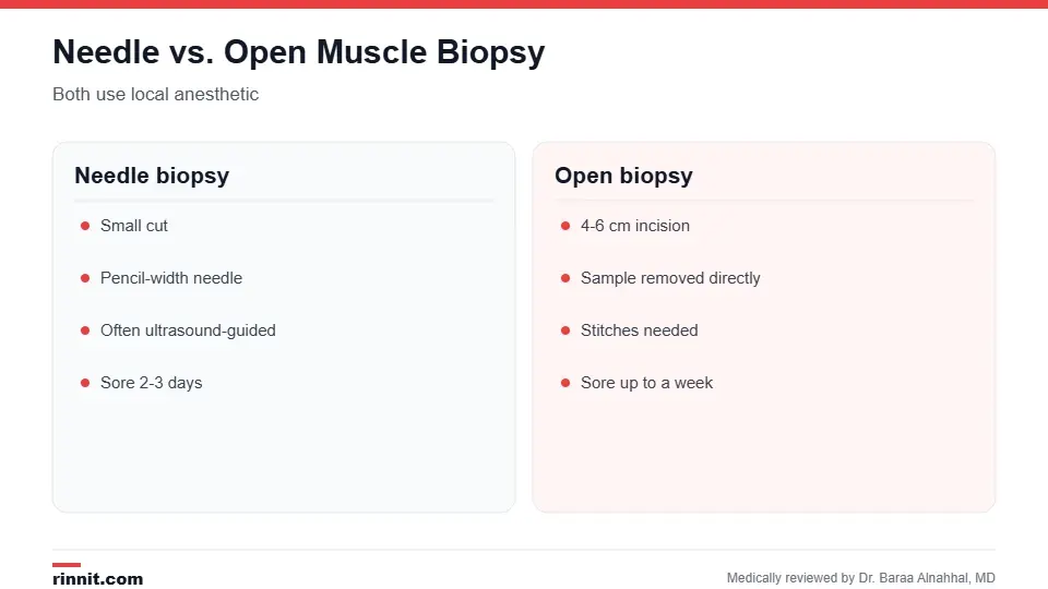
Your provider chooses the method based on your situation.
Type | How it's done | Recovery |
Needle biopsy | Small cut; pencil-width needle, often guided by ultrasound; usually 3 to 4 samples | Sore for 2 to 3 days |
Open biopsy | 4 to 6 cm incision; sample removed; stitches | Sore for up to a week |
Sources: [1]
How to prepare
Tell your provider about muscle injections, muscle tests such as EMG or muscle injuries in the past 6 months.
Stop aspirin or blood thinners only if your provider tells you to.
Fast for several hours if instructed.
Arrange a driver if you'll have sedation or general anesthesia. Sources: [1]
What happens during the procedure

The procedure is usually done as an outpatient.
The skin over the muscle, often the thigh or upper arm, is cleaned.
Local anesthetic numbs the area; sedation or general anesthesia may also be used.
For a needle biopsy, a small cut is made and the needle takes several samples.
For an open biopsy, a larger incision is made and a small piece of muscle is removed.
Pressure, bandages and, for open biopsies, stitches are applied. Sources: [1]
Risks and recovery
Complications are rare but can include infection, a muscle hernia, the wound reopening, scarring or injury to nearby muscles, nerves or blood vessels. Expect soreness and possible bruising for 2 to 3 days, or up to a week after an open biopsy. Call your provider for redness, swelling, warmth, pus or fever. Sources: [1]
Results
Results can take from a few days to more than a week. The report describes the muscle fibers, nerves, inflammation and any abnormal mitochondria, glycogen or fat. Abnormal results often lead to a specific diagnosis and treatment plan. If changes are minimal, the result may be reported as "unspecified myopathy," and another biopsy may be needed. Sources: [1, 2, 3]
Conclusion
A muscle biopsy gives a direct look inside your muscle tissue. It can confirm or clarify the cause of muscle weakness or pain and guide the right treatment, with a quick procedure and a short recovery.
Frequently asked questions
What is a muscle biopsy?
A procedure to remove a small muscle sample for lab testing.
Why is a muscle biopsy done?
To diagnose muscle diseases such as muscular dystrophy or myositis.
Is a muscle biopsy painful?
The area is numbed, but you may feel pressure and soreness afterward.
What's the difference between needle and open biopsy?
A needle biopsy uses a small cut; an open biopsy uses a larger incision.
How long does soreness last?
About 2 to 3 days, or up to a week after an open biopsy.
Do I need to stop blood thinners?
Only if your provider tells you to.
Will I have stitches?
Usually only after an open biopsy.
What are the risks?
Rarely infection, bleeding, scarring or nerve injury.
When will I get results?
From a few days to more than a week.
What is unspecified myopathy?
A result with minimal changes that may need more testing.
Related Reading
EMG (Electromyography): What It Tests, What to Expect and Results a nerve and muscle test often done first.
Creatine Kinase (CK) Test: What It Is, Normal Levels and High CK Causes a blood test for muscle injury.
What Is Multiple Sclerosis? A Plain-Language Guide to Symptoms, Types, and How It's Treated a condition a biopsy can help rule out.
Skin Biopsy: Types, What to Expect, Results and Aftercare another type of tissue sample.
References
Cleveland Clinic. Muscle Biopsy: What It Is, Purpose, Procedure & Recovery. Cleveland Clinic. Last updated February 20, 2024. https://my.clevelandclinic.org/health/diagnostics/muscle-biopsy
Lundberg IE, Tjärnlund A, Bottai M, et al. 2017 European League Against Rheumatism/American College of Rheumatology classification criteria for adult and juvenile idiopathic inflammatory myopathies and their major subgroups. Annals of the Rheumatic Diseases. 2017;76(12):1955-1964. PMID 29079590. https://doi.org/10.1136/annrheumdis-2017-211468
Lai CH, Melli G, Chang YJ, et al. Open muscle biopsy in suspected myopathy: diagnostic yield and clinical utility. European Journal of Neurology. 2010;17(1):136-142. PMID 19674068. https://doi.org/10.1111/j.1468-1331.2009.02765.x
Medical disclaimer
This article is for general education only and does not replace advice from a qualified healthcare professional. Talk to your doctor about your own diagnosis and treatment, and seek emergency care right away for severe or sudden symptoms.



Comments