Knee X-Ray: Why It's Done, What It Shows and What to Expect

Updated: 2 days ago
Written and medically reviewed by Dr. Baraa Alnahhal, MD · Last reviewed: November 2026 · How we review
Quick Answer
A knee X-ray is a painless imaging test that shows the bones of the knee. It's used after injuries and for ongoing knee pain to look for fractures, dislocations, osteoarthritis and bone spurs. It takes 10 minutes and uses very little radiation. Soft tissues such as ligaments and the meniscus don't show clearly, so MRI is used for those.
What the research says
A simple rule decides who needs an X-ray: a meta-analysis of 8 studies and 7,385 adults with acute knee injuries found the Ottawa knee rule had 99% sensitivity and 49% specificity for fractures, meaning it almost never misses a fracture while safely avoiding many unnecessary X-rays (Sims JI et al., European Radiology, 2020) [2]
The finding holds across more studies: a larger meta-analysis of 18 studies and 6,702 patients with knee trauma confirmed a pooled sensitivity of 98% and specificity of 43% for the Ottawa knee rule (Kazemi SM et al., Archives of Academic Emergency Medicine, 2023) [3]
Key takeaways
Shows the patella, femur, tibia and part of the fibula; bone appears white, soft tissue gray.
Finds fractures, dislocations, osteoarthritis, bone spurs, joint fluid, loose fragments, infection and bone tumors.
At least three views: front, side and a kneecap (sunrise) view; about 10 minutes.
Doesn't show ligaments, tendons or the meniscus clearly; MRI or CT may follow.
After an injury, the Ottawa knee rule helps decide whether an X-ray is needed.
What is a knee X-ray?
An X-ray machine sends a small amount of radiation through the knee. Dense bone absorbs more radiation and appears white, while softer tissues appear in shades of gray. A radiologic technologist takes the images, and a radiologist interprets them and sends a report to your provider. Sources: [1]
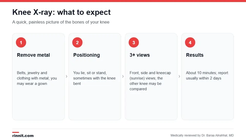
A knee X-ray takes about 10 minutes.
Why is it done?
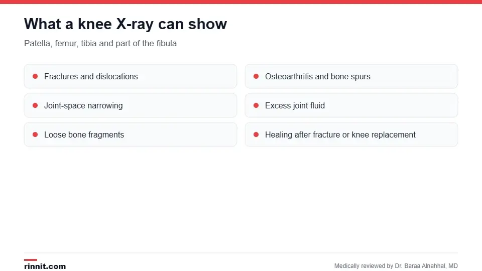
A knee X-ray shows the bones of the knee joint.
Fractures, including a patella fracture, and dislocations.
Knee osteoarthritis, joint-space narrowing and bone spurs.
Excess fluid, which can signal a sprain or other injury.
Loose bone fragments in the joint.
Bone infection (osteomyelitis), bone thinning (osteopenia) or bone tumors.
Misalignment of the joint.
Checking healing after a fracture and monitoring a knee replacement. Sources: [1]
Do you need a knee X-ray after an injury?

The Ottawa knee rule helps decide who needs an X-ray.
Not every knee injury needs an X-ray. Emergency clinicians often use the Ottawa knee rule: an X-ray is recommended if you are 55 or older, have tenderness at the head of the fibula or of the kneecap alone, cannot bend the knee to 90 degrees, or cannot take four steps both right after the injury and in the emergency department. Meta-analyses show this rule misses very few fractures (sensitivity 98–99%). Sources: [2, 3]
How do I prepare?
Wear clothing without metal, and remove jewelry, belts and other metal items, which can hide parts of the image. Tell the technologist if you are or might be pregnant. Sources: [1]
What happens during the X-ray?
You may be given a lead apron to protect the rest of your body; the room may feel cool.
You lie, sit or stand, sometimes with the knee bent, while the technologist positions your leg.
At least three images are taken: front (anteroposterior), side (lateral) and a kneecap view with the knee bent (sunrise view). The other knee may be X-rayed for comparison.
You stay still and may be asked to hold your breath briefly; the technologist steps behind a wall for each image.
Images are checked and blurry ones retaken. Sources: [1]
The test takes about 10 minutes and is painless, though positioning an injured knee may be uncomfortable. Sources: [1]
Understanding your results
Finding | What it may mean |
Break in the bone outline | Fracture |
Narrowed joint space, bone spurs | Osteoarthritis |
Fluid in the joint | Injury such as a sprain, or inflammation |
Loose fragment | Bone or cartilage piece floating in the joint |
Bone thinning or lucent areas | Osteopenia, infection or a bone lesion |
Normal X-ray with ongoing pain | Soft-tissue injury such as a ligament or meniscus tear; MRI may be next |
In an emergency, results are available right away; otherwise the radiologist's report is usually ready within two days. Because X-rays don't clearly show ligaments, tendons or the meniscus, your provider may order an MRI or CT if your symptoms point to a soft-tissue injury. Sources: [1]
Radiation and safety
The radiation dose is very low and usually causes no side effects.
Children are slightly more sensitive, so lower doses may be used.
In pregnancy, a lead apron and other precautions reduce exposure if the X-ray is urgent. Sources: [1]
Conclusion
A knee X-ray is the quick first look at the bones of an injured or painful knee. It reliably shows fractures and arthritis, while the Ottawa knee rule helps avoid unnecessary X-rays after minor injuries. If your X-ray is normal but your knee still hurts, ask whether an MRI is needed to check the ligaments and meniscus.
Keep learning: read about arthritis of the knee and our guide to knee pain.
Frequently asked questions
How long does a knee X-ray take?
About 10 minutes.
How many views are taken?
At least three: front, side and a kneecap (sunrise) view. The other knee is sometimes imaged for comparison.
Can a knee X-ray show a torn meniscus or ACL?
No. These soft tissues don't show clearly on X-rays; an MRI is used instead.
Can a knee X-ray show arthritis?
Yes. Joint-space narrowing and bone spurs are typical signs of osteoarthritis.
Do I need an X-ray after every knee injury?
Not always. The Ottawa knee rule helps decide; it misses very few fractures.
Is a knee X-ray safe?
Yes. The radiation dose is very low, with precautions for children and pregnancy.
Why might I need to stand for the X-ray?
Standing (weight-bearing) views show how much joint space remains under load, which helps assess arthritis.
When will I get my results?
Right away in an emergency; otherwise usually within two days.
What if my X-ray is normal but my knee still hurts?
The problem may be in a ligament, tendon or the meniscus. MRI or further exam may be needed.
Who reads the X-ray?
A radiologist, a doctor trained in imaging, who sends the report to your provider.
Related Reading
What is Knee Osteoarthritis? the most common finding on knee X-rays in adults.
What Is Arthritis of the Knee? Symptoms, Types, and Treatment the types of arthritis that affect the knee.
Patella Fracture: Symptoms, Types, and Recovery Guide a fracture a knee X-ray confirms.
Knee Pain: Symptoms, Causes, Diagnosis, and Treatment Guide the broader work-up of knee pain.
Partial Knee Replacement surgery that X-rays help plan and monitor.
Patellar Grind Test (Clarke's Sign): Checking Kneecap Pain explains the knee exam that checks for cartilage problems under the kneecap.
References
Cleveland Clinic. Knee X-Ray: Anatomy, Procedure & What to Expect. Cleveland Clinic. Last updated July 18, 2022. https://my.clevelandclinic.org/health/diagnostics/23501-knee-x-ray
Sims JI, Chau MT, Davies JR. Diagnostic accuracy of the Ottawa Knee Rule in adult acute knee injuries: a systematic review and meta-analysis. European Radiology. 2020;30(8):4438-4446. PMID 32222797. https://doi.org/10.1007/s00330-020-06804-x
Kazemi SM, Khorram R, Fayyazishishavan E, et al. Diagnostic accuracy of Ottawa Knee Rule for diagnosis of fracture in patients with knee trauma: a systematic review and meta-analysis. Archives of Academic Emergency Medicine. 2023;11(1):e30. PMID 37215241. https://doi.org/10.22037/aaem.v11i1.1934
Medical disclaimer
This article is for general education only and does not replace advice from a qualified healthcare professional. Talk to your doctor about your own diagnosis and treatment, and seek emergency care right away for severe or sudden symptoms.



Comments