Hand X-Ray: What It Shows, How It's Done and What Results Mean

Updated: 2 days ago
Written and medically reviewed by Dr. Baraa Alnahhal, MD · Last reviewed: November 2026 · How we review
Quick Answer: What is a hand X-ray?
A hand X-ray (radiograph) is a fast, painless imaging test that makes a black-and-white picture of the inside of the hand. It helps providers find broken bones, foreign objects, arthritis, bone tumors and abnormal bone growth. The test takes 5 to 10 minutes, uses a very small dose of radiation, and results are ready within one to two days. [1]
What the research says
Standard X-rays can miss some small wrist bone fractures. In a meta-analysis of 7 studies with 3,373 X-ray images, AI software found scaphoid fractures with a pooled sensitivity of 80% and specificity of 89%. The authors note these fractures are easy to miss on X-rays and often hidden at first. (Kraus et al., European Radiology, 2024) [2]
A low-radiation 3D scan can help when X-rays look normal. Across 5 studies with 439 patients, cone-beam CT detected scaphoid fractures with 87.7% sensitivity and 99.2% specificity, and other carpal fractures with 90.6% sensitivity. (Fitzpatrick et al., Skeletal Radiology, 2022) [3]
Key takeaways
A hand X-ray shows the bones of your hand in black and white; calcium-rich bone appears white. [1]
It helps diagnose fractures, foreign objects, arthritis, bone tumors and abnormal bone growth. [1]
Children may have a hand X-ray to check "bone age" and growth. [1]
The test is quick and painless, and the radiation dose is minimal. [1]
Tiny fractures can be missed where bones overlap, so a CT scan or MRI may follow if pain continues. [1]
What is a hand X-ray?
A hand X-ray is a test that creates a picture of the structures inside your hand. X-rays are a form of radiation made of electromagnetic waves. They pass through your body and form an image on special film or a digital detector. [1]
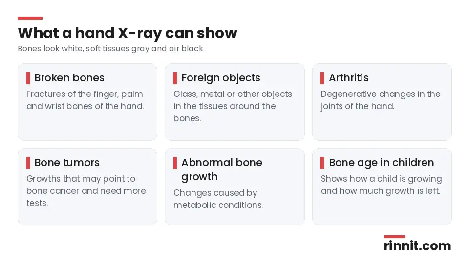
Different tissues absorb different amounts of radiation. Bones are dense and full of calcium, so they absorb more and look white. Muscle, fat and other soft tissues let more radiation through and appear in shades of gray, while air looks black. [1]
X-rays are the oldest type of medical imaging still in use. They were discovered in 1895, first used to picture human tissue in 1896, and are still the most commonly used imaging test today. [1]
Why would you need a hand X-ray?
Your provider may order a hand X-ray to look for the cause of pain, swelling or an injury in one or both hands. It can help diagnose: [1]
What it can show | What it means |
Broken bones (fractures) | A crack or break in one of the bones of your fingers, palm or wrist |
Foreign objects | Glass, metal or other objects in the soft tissues around your hand bones |
Degenerative changes | Signs of arthritis in the joints of your hand |
Tumors | Bone growths that may point to bone cancer and need more testing |
Abnormal bone growth | Bone changes caused by metabolic conditions |
Sources: [1]
Bone age in children
A pediatrician may request an X-ray of a child's hand to estimate their "bone age." This helps show whether a health problem is affecting normal growth and how much growing the child has left. [1]
Who performs a hand X-ray?
A radiologic technologist (X-ray technician) performs the test. These professionals are trained in patient care, radiation protection, positioning and imaging procedures. A doctor called a radiologist then reads the images. [1]
How do you prepare for a hand X-ray?
Very little preparation is needed. Before the test: [1]
Remove all rings, bracelets, watches and other jewelry from your hand and wrist, because metal can block parts of the image.
Tell the technologist if you are pregnant or could be pregnant.
Ask any questions you have; the technologist will explain the steps first.
The radiation dose from a hand X-ray is minimal and considered safe during pregnancy, but your provider will decide whether the X-ray is needed, and the technologist will take steps to reduce exposure to the fetus. [1]
What happens during a hand X-ray?
The test is done in a provider's office or a hospital radiology department. You may be given a lead apron to shield your reproductive organs. The room may feel cool, but the whole test usually takes only 5 to 10 minutes, and you will not feel the radiation. [1]
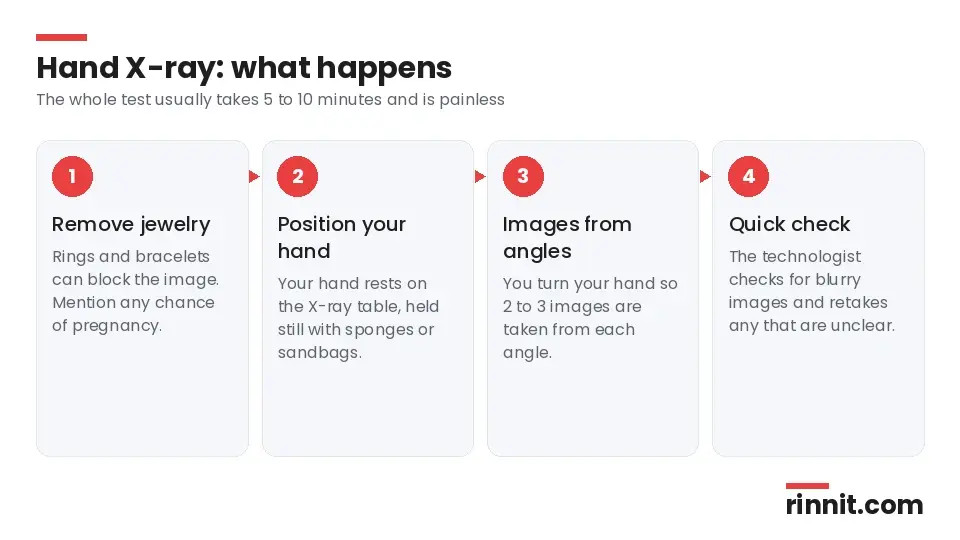
You place your hand on the X-ray table. Sponges, sandbags, pillows or other supports may hold your arm still.
The technologist places a film holder or digital plate under the table, then steps behind a wall or into a small room to run the machine.
You may be asked to turn your hand into different positions, and sometimes to hold a weight, so the images show your hand from several angles.
Two or three images are usually taken from each angle. Keep very still, because movement blurs the pictures; you may be asked to hold your breath for a few seconds.
Let the technologist know if positioning your hand is painful. They can help you through the test. [1]
What happens after a hand X-ray?
The technologist may ask you to wait a few minutes while they check that no image is blurry, and they will retake any that are. A radiologist then reviews the images and sends a report to your provider, who will go over the findings and recommend treatment. [1]
Depending on the results, you may be asked to come back for more views of your hand or for repeat X-rays to see how a condition changes over time. [1]
When will you get your results?
If the X-ray was done in an emergency, results may be available almost right away. Otherwise, the radiologist usually has results ready in one to two days. [1]
Can a hand X-ray miss a fracture?
Yes. Sometimes a broken hand bone does not show on the first X-ray. These missed fractures are usually tiny and sit where bones overlap. If your X-ray looks normal but you still have pain and tenderness, your provider may order a CT scan or an MRI. [1] Studies show that wrist bone fractures in particular are easy to miss on standard X-rays, and newer tools such as cone-beam CT are being used to find them. [2] [3]

What are the risks of a hand X-ray?
Hand X-rays rarely cause side effects. The radiation dose is minimal and passes straight through your body. [1]
Pregnancy: there is a slightly higher risk from radiation exposure, so always tell the technologist. A lead apron may be used. [1]
Children: there is also a slightly higher risk, so lower doses may be used. [1]
Cancer risk: heavy radiation exposure carries a slight risk of cancer, but the benefit of an accurate diagnosis outweighs the small risk from a hand X-ray. Ask your provider if you are concerned. [1]
Conclusion
A hand X-ray is one of the simplest ways to see what is going on inside your hand. It takes minutes, does not hurt and uses very little radiation, yet it can confirm a fracture, find a foreign object or show signs of arthritis. [1]
If your pain continues after a normal X-ray, go back to your provider. Some fractures only show on more detailed scans, and catching them early helps your hand heal properly.
Keep learning: Read our guide to X-rays or our article on boxer's fracture. For more background, see MedlinePlus: Hand x-ray.
FAQ
What is a hand X-ray?
A quick, painless imaging test that makes a black-and-white picture of the bones and soft tissues inside your hand. [1]
What can a hand X-ray show?
Broken bones, foreign objects, arthritis, bone tumors and abnormal bone growth from metabolic conditions. [1]
Does a hand X-ray hurt?
No. You will not feel the radiation, though holding some positions may be uncomfortable if your hand is injured. [1]
How long does a hand X-ray take?
Usually about 5 to 10 minutes. [1]
How do I prepare for a hand X-ray?
Remove jewelry from your hand and wrist, and tell the technologist if you are or might be pregnant. [1]
Is a hand X-ray safe during pregnancy?
The dose is minimal and considered safe, but your provider decides if it is needed and precautions are taken to protect the fetus. [1]
How soon will I get my results?
Almost right away in an emergency; otherwise usually within one to two days. [1]
Can a hand X-ray miss a fracture?
Yes. Tiny fractures where bones overlap can be missed, so a CT scan or MRI may be ordered if pain continues. [1]
Why do children get hand X-rays for bone age?
Bone age helps a pediatrician see whether a child is growing normally and how much growth is left. [1]
Who reads my hand X-ray?
A radiologist reviews the images and sends a report to your provider, who explains the results. [1]
Related Reading
Boxer's Fracture: Symptoms, Treatment, and Recovery Time Explained explains a common hand fracture that a hand X-ray confirms.
Understanding Metacarpal Fractures: Symptoms, Types, and Recovery covers breaks in the long bones of the palm and how they are found on X-ray.
Rheumatoid Arthritis — Symptoms, Causes, Diagnosis, Treatment & Complete Guide describes a joint disease whose hand changes can show on X-ray.
Gout in Hands, Fingers, and Wrists explains gout in the small joints that a hand X-ray can help assess.
References
Cleveland Clinic. Hand X-Ray: Anatomy, Procedure & What To Expect. Cleveland Clinic. Last updated July 18, 2022. https://my.clevelandclinic.org/health/diagnostics/23518-hand-x-ray
Kraus M, Anteby R, Konen E, et al. Artificial intelligence for X-ray scaphoid fracture detection: a systematic review and diagnostic test accuracy meta-analysis. Eur Radiol. 2024;34(7):4341-4351. PMID 38097728. https://doi.org/10.1007/s00330-023-10473-x
Fitzpatrick E, Sharma V, Rojoa D, et al. The use of cone-beam computed tomography (CBCT) in radiocarpal fractures: a diagnostic test accuracy meta-analysis. Skeletal Radiol. 2022;51(5):923-934. PMID 34542681. https://doi.org/10.1007/s00256-021-03883-9
Medical disclaimer
This article is for general education only and does not replace advice from a qualified healthcare professional. Talk to your doctor about your own diagnosis and treatment, and seek emergency care right away for severe or sudden symptoms.



Comments