Electrophysiology (EP) Study: Why It's Done, What Happens and Recovery

Updated: 2 days ago
Written and medically reviewed by Dr. Baraa Alnahhal, MD · Last reviewed: November 2026 · How we review
Quick Answer
An electrophysiology (EP) study is a procedure that records the heart's electrical activity from inside the heart to diagnose abnormal heart rhythms (arrhythmias). Thin catheters are threaded through a vein to the heart. If a treatable cause is found, catheter ablation is often done in the same session. Most people resume normal activities the next day.
What the research says
An EP study can change the diagnosis: in 22 patients referred for a defibrillator because their fast heart rhythm looked like ventricular tachycardia, the EP study showed a less dangerous supraventricular rhythm in every case; 95% were successfully treated with ablation and 82% avoided needing a defibrillator (William J et al., Journal of Electrocardiology, 2024) [2]
It helps sort out who is at risk: among 143 adults having ventricular stimulation during an EP study for fainting, nonsustained ventricular tachycardia or defibrillator decisions, 23.8% had an inducible ventricular arrhythmia, and those patients had weaker heart pumping (ejection fraction 38% vs 50%) (Isaza N et al., Heart Rhythm, 2024) [3]
Key takeaways
Records electrical signals from inside the heart to find the type and source of an arrhythmia.
Used when an EKG, Holter or event monitor doesn't give enough information.
No food or drink after midnight; arrange a driver; ask which medicines to stop.
Catheters go through a vein in the groin, arm or neck; ablation can be done at the same time.
Serious complications are uncommon; the risk of death is about 1 in 5,000.
What is an EP study?
Your heartbeat is controlled by electrical signals that travel through a specialized conduction system. Surface tests like an EKG record these signals from the skin, but an EP study places electrodes inside the heart chambers, giving a far more detailed map. This shows where an abnormal rhythm starts, how it travels and how best to treat it. Sources: [1]
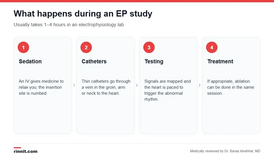
An EP study usually takes one to four hours.
Why is it done?

Reasons your provider may recommend an EP study.
To identify the type, cause and origin of an arrhythmia, such as supraventricular tachycardia, Wolff-Parkinson-White syndrome or ventricular tachycardia.
To find the cause of unexplained dizziness or fainting.
To assess your risk of sudden cardiac arrest.
To check how well a heart rhythm medicine works.
To decide on treatment such as ablation, a pacemaker or an implantable cardioverter-defibrillator (ICD). Sources: [1]
It's usually ordered when other tests, such as an EKG, Holter monitor, event recorder, stress test or echocardiogram, haven't given a clear answer. Sources: [1]
How do I prepare?
Arrange for someone to drive you home; you can't drive for at least 24 hours.
Eat a normal meal the evening before, then nothing by mouth after midnight (including water, gum and mints), except small sips of water with approved medicines.
Ask which medicines to stop or continue, and when to restart them; some rhythm medicines are paused beforehand.
Ask about adjusting diabetes medicines.
Tell your team about latex allergies or past problems with anesthesia, and bring a one-day supply of your medicines. Sources: [1]
What happens during the study?
An IV is placed in your arm or hand for fluids and medicines that relax you; you're drowsy but not asleep.
The insertion site, usually the groin, is cleaned and numbed.
Several catheters are threaded through a vein to the heart, guided by X-ray (fluoroscopy) and ultrasound.
The catheters record electrical signals and map the conduction system.
Small electrical pulses pace the heart, and medicines such as isoproterenol or adenosine may be used to reproduce the abnormal rhythm.
If appropriate, catheter ablation treats the faulty pathway in the same session.
If a dangerous rhythm occurs, a shock through chest patches restores a normal rhythm. Sources: [1]
You shouldn't feel pain, though you may feel pressure where the catheters go in, or a racing heart when the rhythm is triggered. The study takes one to four hours. Sources: [1]
Recovery
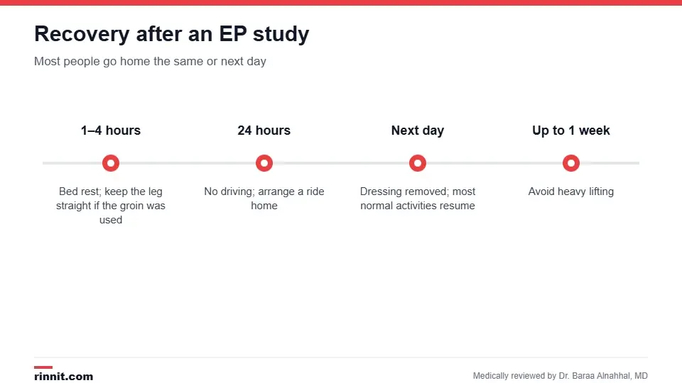
Most people resume normal activities the next day.
Stay in bed for one to four hours; keep your leg straight if the groin was used.
Most people don't need stitches; the dressing is removed the next day.
Keep the site clean and dry; you can eat and take medicines afterward.
Resume most normal activities the next day, but avoid heavy lifting for up to a week. Sources: [1]
Risks
EP studies are generally safe, but complications can include bleeding or infection at the insertion site, an abnormal heart rhythm, blood clots, damage to a blood vessel, valve or heart chamber, and rarely heart attack or stroke. The risk of death is about 1 in 5,000. Sources: [1]
Understanding your results
Preliminary results are available right after the procedure and decide whether you go home or stay in the hospital. Depending on the findings, your options may include medicine, ablation, a pacemaker, an ICD or, rarely, surgery. Some people still need another treatment after ablation. Sources: [1, 2, 3]
Call your provider for redness, swelling or drainage at the site, numbness or tingling in the leg or arm, a cold or discolored hand or foot, vomiting or chest pain. Call 911 for bleeding that doesn't stop with pressure or swelling that quickly gets worse. Sources: [1]
Conclusion
An EP study gives the most detailed picture of your heart's electrical system, turning a vague diagnosis like "palpitations" or "fainting" into a specific rhythm with a specific treatment, often fixed during the same procedure. Ask your cardiologist what they hope to find and whether ablation might be done at the same time.
Keep learning: read about heart arrhythmias and catheter ablation.
Frequently asked questions
What is an EP study used for?
To diagnose the type and source of abnormal heart rhythms and guide treatment.
Is an EP study painful?
No. You're sedated and the site is numbed, though you may feel pressure.
Am I awake during the procedure?
Usually you're drowsy but not fully asleep.
How long does it take?
One to four hours.
Can treatment be done at the same time?
Yes. Catheter ablation is often done during the same session.
Do I need to fast?
Yes. Nothing by mouth after midnight, except small sips of water with approved medicines.
When can I drive?
Not for at least 24 hours.
What are the risks?
Bleeding, infection, blood vessel damage, clots and, rarely, heart attack, stroke or death (about 1 in 5,000).
When can I go back to normal activities?
Usually the next day, avoiding heavy lifting for up to a week.
Will I need to stay in the hospital?
Sometimes, depending on the preliminary results.
Related Reading
Heart Arrhythmia: Symptoms, Types, Causes, Diagnosis & Treatment — Complete Guide the rhythm problems an EP study diagnoses.
Catheter Ablation: How It Works, Success Rates, Risks and Recovery treatment often done during the EP study.
Wolff-Parkinson-White Syndrome Guide: Symptoms, Causes, Diagnosis, and Treatment an extra electrical pathway an EP study can find.
What Is Ventricular Tachycardia? A Plain-Language Guide to the Fast Heart Rhythm That Starts in the Heart's Lower Chambers a dangerous rhythm assessed with an EP study.
Sudden Cardiac Arrest: Symptoms, Causes, Diagnosis, and Treatment Explained the risk an EP study can help estimate.
References
Cleveland Clinic. Electrophysiology (EP) Study: What It Is, What It's Used For. Cleveland Clinic. Last updated November 21, 2023. https://my.clevelandclinic.org/health/diagnostics/23054-electrophysiology-study
William J, Kistler PM, Kalman JM, et al. Aberrancy masquerading as ventricular tachycardia: importance of invasive electrophysiology study for diagnosis of wide complex tachycardias. Journal of Electrocardiology. 2024;85:50-57. PMID 38852223. https://doi.org/10.1016/j.jelectrocard.2024.05.099
Isaza N, Stabenau HF, Kramer DB, et al. The spatial ventricular gradient is associated with inducibility of ventricular arrhythmias during electrophysiology study. Heart Rhythm. 2024;21(11):2160-2167. PMID 38718942. https://doi.org/10.1016/j.hrthm.2024.05.005
Medical disclaimer
This article is for general education only and does not replace advice from a qualified healthcare professional. Talk to your doctor about your own diagnosis and treatment, and seek emergency care right away for severe or sudden symptoms.



Comments