Cellular Dermatofibroma: What This Common, Noncancerous Skin Bump Means for You

Updated: 3 days ago
Medically reviewed by Dr. Baraa Alnahhal, MD · Last reviewed: October 17, 2026
Quick answer
A cellular dermatofibroma is a noncancerous skin growth that appears as a firm bump, often on the legs, and often grows down into the deepest layer of skin. [1] It rarely causes harmful symptoms and often follows an injury at the site. [1] Usually no treatment is needed; if wanted, removal is a short in-office procedure. [1]
What the research says
According to PubMed:
The cellular type is uncommon. In a study of 192 dermatofibromas from 181 patients, 80% were the common type and only 2.1% were the cellular variant; most were on the limbs (74% of patients), and women were more often affected. (Alves et al., Anais Brasileiros de Dermatologia, 2014) [3].
Cellular types can recur, and rarely spread. In 27 cases of cellular fibrous histiocytoma, follow-up of 19 patients showed local recurrence in 5 and, in 1 patient, lung metastases after repeated recurrences. (Zhong and Wang, Zhonghua Bing Li Xue Za Zhi, 2013) [5].
Deeper growth raised the risk of recurrence. In a cohort of 2,858 dermatofibromas, 39% of the 23 facial lesions were the cellular type and 44% had subcutaneous involvement; recurrence was 22% with no metastases. A review of 10 studies of facial dermatofibromas found a 14% pooled local recurrence rate, and facial lesions with subcutaneous involvement were more likely to recur (odds ratio 4.53, 95% CI 1.12–18.24). (Alsawas et al., American Journal of Dermatopathology, 2022) [6].
What is a cellular dermatofibroma?
Cellular dermatofibromas are a type of dermatofibroma — a noncancerous tumor. They appear as firm bumps (nodules), often on your legs. [1] A cellular dermatofibroma may look like a small, firm bump, similar to a mole. [1]
The defining difference is depth. Cellular dermatofibromas extend deeper into your skin's layers than other dermatofibromas. [1] Usually, dermatofibromas are superficial, meaning they grow in the dermis, just under the top layer of skin (the epidermis). Cellular dermatofibromas often grow down to subcutaneous tissue, the deepest layer of your skin. [1] [2]
Compared to other dermatofibromas, cellular dermatofibromas are more likely to come back (recur) after treatment. [1] [2] In about 1 in 10 people, cellular dermatofibromas cause necrosis in the tumor — tissue or cell death in a certain area of your body. [1] [2]

Are cellular dermatofibromas malignant?
By definition, cellular dermatofibromas aren't malignant (noncancerous). But they're more likely than other types of dermatofibromas to spread to other parts of your body (metastasize). [1]
Who might get a cellular dermatofibroma?
Anyone can get a cellular dermatofibroma. Dermatofibromas are most common in people ages 20 to 49, and twice as many women as men have them. [1]
How common are they?
Dermatofibromas are somewhat common. About 3 in every 100 skin lesions is a dermatofibroma. [1] A skin lesion is a growth, mole, or sore on or in your skin.

What causes a cellular dermatofibroma?
About 1 in 5 people who have a dermatofibroma have had an injury or wound where the tumor forms. [1] Sometimes, a dermatofibroma develops spontaneously, for no known reason. [1]
People with conditions that affect their immune system may have multiple dermatofibromas in a cluster. [1] You're more likely to have a dermatofibroma cluster if you have:
HIV/AIDS
Leukemia
Lupus
What are the symptoms of a cellular dermatofibroma?
Cellular dermatofibromas look like small round or oval bumps. They're usually less than 1 centimeter (cm) in diameter. [1] They may be flesh-colored, brown, or reddish-brown. [1]
Usually, dermatofibromas form on your arms or legs. [1] They may feel firm to the touch and can be itchy, tender, or painful. [1] Sometimes, they're shaped like a dimple — the center of the lesion falls in, and the edges of the lesion pucker around it. [1]
How is a cellular dermatofibroma diagnosed?
To diagnose a cellular dermatofibroma, your healthcare provider starts by looking at the lesion. You may have a skin biopsy to confirm whether it's a dermatofibroma or another type of skin lesion. [1]
In a skin biopsy, your healthcare provider removes a small tissue sample and sends it to a lab. In the lab, a pathologist (a doctor who specializes in examining body tissues) views it under a microscope. [1]
Cellular dermatofibroma vs. mole: what's the difference?
You may not be able to tell the difference between a dermatofibroma and a mole at first glance. Cellular dermatofibromas may attach to your subcutaneous tissue. The pinch test helps: if you gently pinch the skin growth, a dermatofibroma usually creates a dimple or indent. [1] Moles stay raised above your skin's surface because they don't attach to the subcutaneous layer. [1]
How is a cellular dermatofibroma treated?
Because they're noncancerous, dermatofibromas don't always require treatment. [1]
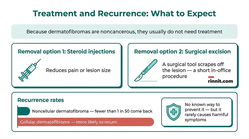
If a dermatofibroma is large or causes discomfort, your healthcare provider may remove it. [1] Removal is a short in-office procedure. [1] Your provider may use:
Removal option | What it does |
|---|---|
Steroid injections | Reduce pain or lesion size [1] |
Surgical excision | The bump is cut out with a scalpel and the skin is stitched closed |
What is the outlook for cellular dermatofibroma, and does it recur?
Cellular dermatofibromas are more likely to return (recur) after treatment than other dermatofibroma types. [1] [2] Other types of dermatofibromas don't usually return after treatment — fewer than 1 in 50 people have a noncellular dermatofibroma come back after removal. [1]
Can I prevent a cellular dermatofibroma?
Cellular dermatofibromas cannot be prevented, since there isn't a known way to stop one from forming. [1] Fortunately, dermatofibromas aren't cancerous. If you get one, it rarely causes discomfort or harmful symptoms.
What questions should I ask my doctor?
You may want to ask your healthcare provider about the cause of a cellular dermatofibroma, whether you need treatment and the risks and benefits of removing it.
What's the most likely cause of a cellular dermatofibroma?
Is a skin growth a dermatofibroma or another condition?
Do I need treatment for a dermatofibroma?
What are the benefits and risks of removing a dermatofibroma?
Is there anything I can do to prevent a dermatofibroma from returning after treatment?
When should you see a provider about a cellular dermatofibroma?
Most dermatofibromas are harmless and need no treatment. It's still wise to have any new, changing, or unexplained skin growth checked by a healthcare provider — the pinch test, a visual exam, and (if needed) a biopsy are the standard way to confirm what a bump is. If a bump is itchy, tender, painful, growing, or bleeding, mention it at your next appointment.
What's the bottom line?
A cellular dermatofibroma is a small, firm, noncancerous skin bump — usually under 1 centimeter, most often on the arms or legs — that differs from ordinary dermatofibromas because it extends down to the skin's deepest layer. It most often shows up in people ages 20 to 49 (twice as many women as men), sometimes forms after an injury at the site, and is diagnosed with a simple visual exam plus, when needed, a skin biopsy. Because it's benign, treatment is usually unnecessary; removal is a quick in-office procedure reserved for bumps that cause discomfort. The one caveat: cellular dermatofibromas are more likely to recur after removal than other types, so follow-up care is worth discussing with your provider.
Ready to learn more? Visit Rinnit.com for trusted, easy-to-understand health guides like this one.
Conclusion
Discovering a new bump on your skin is naturally unsettling, but a cellular dermatofibroma is among the more reassuring explanations — a noncancerous growth that most often needs nothing more than a watchful eye. Knowing how it differs from a mole (the pinch test), what depth means for the cellular type, and how recurrence works after removal puts you in control of the conversation with your provider. When in doubt, a quick skin check is the simplest step.
Find more compassionate, plain-English health guides at Rinnit.com.
Frequently Asked Questions
What is a cellular dermatofibroma?
A cellular dermatofibroma is a noncancerous skin growth — a type of dermatofibroma (noncancerous tumor) — that appears as a firm bump (nodule), often on the legs. It looks like a small, firm bump similar to a mole.
What does "cellular" mean in cellular dermatofibroma?
The "cellular" type differs in depth. While ordinary dermatofibromas stay in the dermis, just under the skin's top layer, cellular dermatofibromas often extend down to the subcutaneous tissue, the deepest layer of your skin.
Is a cellular dermatofibroma cancerous?
No. By definition, cellular dermatofibromas aren't malignant — they are noncancerous.
Can cellular dermatofibromas spread?
Cellular dermatofibromas are more likely than other types of dermatofibromas to spread to other parts of your body (metastasize), even though they are noncancerous.
How common are dermatofibromas?
Dermatofibromas are somewhat common — about 3 in every 100 skin lesions is a dermatofibroma. A skin lesion is any growth, mole, or sore on or in your skin.
Who is most likely to get one?
Anyone can get one. Dermatofibromas are most common in people ages 20 to 49, and twice as many women as men have them.
What causes a cellular dermatofibroma?
About 1 in 5 people who have a dermatofibroma have had an injury or wound where the tumor forms. Sometimes it develops spontaneously, for no known reason.
Can immune system conditions cause more dermatofibromas?
Yes. People with conditions that affect the immune system may have multiple dermatofibromas in a cluster. This is more likely if you have HIV/AIDS, leukemia, or lupus.
What does a cellular dermatofibroma look like?
A cellular dermatofibroma looks like a small round or oval bump, usually less than 1 centimeter in diameter. It may be flesh-colored, brown, or reddish-brown.
Where do dermatofibromas usually appear?
Dermatofibromas usually form on your arms or legs.
Do dermatofibromas hurt?
Dermatofibromas may feel firm to the touch and can be itchy, tender, or painful.
What is the dimple shape?
Sometimes a dermatofibroma is shaped like a dimple — the center of the lesion falls in, and the edges of the lesion pucker around it.
How is a cellular dermatofibroma diagnosed?
Your provider starts by looking at the lesion. You may have a skin biopsy to confirm whether it's a dermatofibroma or another type of skin lesion. In a biopsy, a small tissue sample is removed and sent to a lab, where a pathologist (a doctor who specializes in examining body tissues) views it under a microscope.
How do I tell a dermatofibroma from a mole?
It can be hard at first glance, but the pinch test helps: if you gently pinch the skin growth, a dermatofibroma usually creates a dimple or indent. Moles stay raised above the skin's surface because they don't attach to the subcutaneous layer.
Does a cellular dermatofibroma need treatment?
Usually not. Because dermatofibromas are noncancerous, they don't always require treatment.
When is removal recommended?
If a dermatofibroma is large or causes discomfort, your provider may remove it. Removal is a short in-office procedure.
How is a dermatofibroma removed?
Providers may use steroid injections to reduce pain or lesion size, or surgical excision, using a surgical tool to scrape off the lesion.
What is necrosis?
Necrosis is tissue or cell death in a certain area of your body. In about 1 in 10 people, cellular dermatofibromas cause necrosis in the tumor.
Will it come back after removal?
Cellular dermatofibromas are more likely to return (recur) after treatment than other dermatofibroma types. Other types don't usually return — fewer than 1 in 50 people have a noncellular dermatofibroma come back after removal.
Can I prevent a cellular dermatofibroma?
There isn't a known way to prevent one. Fortunately, dermatofibromas aren't cancerous, and if you get one, it rarely causes discomfort or harmful symptoms.
What questions should I ask my doctor?
Consider asking about the most likely cause, whether the growth is a dermatofibroma or another condition, whether you need treatment, the benefits and risks of removal, and anything you can do to prevent recurrence.
What is the subcutaneous tissue?
The subcutaneous tissue is the deepest layer of your skin (also called the hypodermis). Cellular dermatofibromas often grow down into this layer, unlike superficial dermatofibromas that stay in the dermis.
When should I have a skin bump checked?
Any new, changing, or unexplained skin growth is worth having a healthcare provider look at. Mention it promptly if a bump is itchy, tender, painful, growing, or bleeding.
This article is for general informational purposes only and does not constitute medical advice, diagnosis, or treatment. Always seek the guidance of your physician or other qualified health provider with any questions about a medical condition. Never disregard professional medical advice or delay seeking care because of something you have read here. Any new or changing skin growth should be evaluated by a qualified healthcare provider.
Related Reading
Dermatofibrosarcoma Protuberans (DFSP): Symptoms, Causes, Diagnosis & Treatment — Complete Guide explains the rare skin cancer doctors rule out when checking these bumps.
Skin Biopsy: Types, What to Expect, Results and Aftercare describes how a small skin sample confirms what a bump is.
Lipoma: Symptoms, Causes, Diagnosis, and Treatment covers soft fatty lumps under the skin and when to remove them.
Skin Tags (Acrochordons): Understanding Common Skin Growths explains another common harmless skin growth and its removal.
References
Cleveland Clinic. Cellular Dermatofibroma. Cleveland Clinic. Last updated April 8, 2022. https://my.clevelandclinic.org/health/diseases/22668-cellular-dermatofibroma
Cheng H, Emanuel P. Dermatofibroma (fibrous histiocytoma) pathology. DermNet. 2013. https://dermnetnz.org/topics/dermatofibroma-fibrous-histiocytoma-pathology
Alves JV, Matos DM, Barreiros HF, et al. Variants of dermatofibroma — a histopathological study. An Bras Dermatol. 2014;89(3):472-477. PMID 24937822. https://doi.org/10.1590/abd1806-4841.20142629
Elbendary A, Griffin JR, Elston DM, et al. Cellular dermatofibroma: a hyperkeratotic indurated plaque on the thigh. Indian Dermatol Online J. 2016;7(4):308-310. PMID 27559511. https://doi.org/10.4103/2229-5178.185497
Zhong YP, Wang J. Cellular fibrous histiocytoma: a clinicopathologic analysis of 27 cases [article in Chinese]. Zhonghua Bing Li Xue Za Zhi. 2013;42(3):153-157. PMID 23769432. https://doi.org/10.3760/cma.j.issn.0529-5807.2013.03.003
Alsawas M, Dundar B, Prokop L, et al. Clinicopathologic features of facial dermatofibromas, a cohort study, and a systematic review of literature. Am J Dermatopathol. 2022;44(11):822-827. PMID 35503883. https://doi.org/10.1097/DAD.0000000000002211



Comments