Auscultation: How Doctors Listen to Your Heart, Lungs and Belly and What the Sounds Mean

Updated: 2 days ago
Written and medically reviewed by Dr. Baraa Alnahhal, MD · Last reviewed: November 2026 · How we review
Quick Answer: What is auscultation?
Auscultation is when a provider listens to the sounds of your heart, lungs, arteries and belly with a stethoscope. It's a routine, safe, non-invasive part of a physical exam. Your provider listens for the timing, loudness, pitch and quality of each sound, and unusual sounds such as murmurs, wheezes or crackles can point to conditions that need more testing. [1]
What the research says
Hearing a third heart sound strongly points to heart failure. In a review of studies in emergency patients with shortness of breath, an S3 heard on auscultation raised the likelihood of acute heart failure about fourfold (positive likelihood ratio 4.0). (Martindale et al., Academic Emergency Medicine, 2016) [2]
Careful listening can rule in valve disease with few false alarms. In 175 people referred for a heart murmur, auscultation correctly picked up 83% to 91% of valve problems later confirmed by echocardiogram, with 100% specificity. (Draper et al., Heart, 2019) [3]
Digital stethoscopes with AI are being tested in children. Across 41 studies, computer models detected wheezing with a pooled sensitivity of about 90% and specificity of about 96%, though most used small, single-center data. (Park et al., Journal of Medical Internet Research, 2025) [4]
Key takeaways
Auscultation means listening to body sounds with a stethoscope during a physical exam. [1]
Providers listen to four areas: the heart, lungs, arteries and abdomen. [1]
Normal heart sounds have a steady "lub-dub" rhythm, called S1 and S2. [1]
Abnormal sounds such as murmurs, wheezes, crackles or absent bowel sounds can signal a problem. [1]
An unusual finding usually leads to more tests, not an instant diagnosis. [1]
What is auscultation?
Auscultation is listening to the sounds your heart, lungs, arteries and belly make. Your provider places a stethoscope directly on your chest, neck, back or abdomen and listens for different sounds, along with their quality, volume and timing. [1]
It is one of the oldest techniques in medicine. It is safe, easy and non-invasive, and it helps providers spot possible medical conditions early. [1]
When is auscultation done?
Your provider uses auscultation during routine physical examinations to check your circulatory, respiratory and gastrointestinal systems. They may use it to keep an eye on your overall health or to confirm or rule out a specific condition. [1]
What are the types of auscultation?

A stethoscope exam can cover four body areas.
Type | Where the stethoscope goes | What your provider listens for |
Heart (cardiac) | Five points on the upper left chest | Timing, intensity, pitch and length of heartbeats |
Lungs (pulmonary) | Front and back of the chest, top to bottom | Length of breaths, pitch, loudness and abnormal sounds such as wheezing |
Arteries | Neck, abdomen and over the kidneys | Bruits (rough blood flow) and weak or absent pulses |
Abdomen (bowel) | Different regions of the belly | Whether bowel sounds are present, how often and how they sound |
Sources: [1]
Heart auscultation
You sit up or lie back at about a 45-degree angle. Your provider may ask you to turn toward your left side so they can hear your heart more clearly. They move the stethoscope across five points on your chest where the heart valves are heard best. [1]
Lung auscultation
You sit upright if you can and breathe deeply through your mouth. Your provider listens from the top of your lungs downward, first on the front of your chest and then on your back, hearing one full breath at each spot and comparing the two sides. [1]
Artery auscultation
Your provider can listen over the arteries in your neck, abdomen and kidneys. A whooshing sound called a bruit suggests rough, uneven blood flow, which can mean poor circulation. A weak or absent pulse can point to a blocked artery. [1]
Abdominal auscultation
You lie on your back with your head on a pillow, sometimes with a support under your knees. Your provider moves the stethoscope over different parts of your belly to hear the sounds your bowels make. [1]
What are the five auscultation points of the heart?
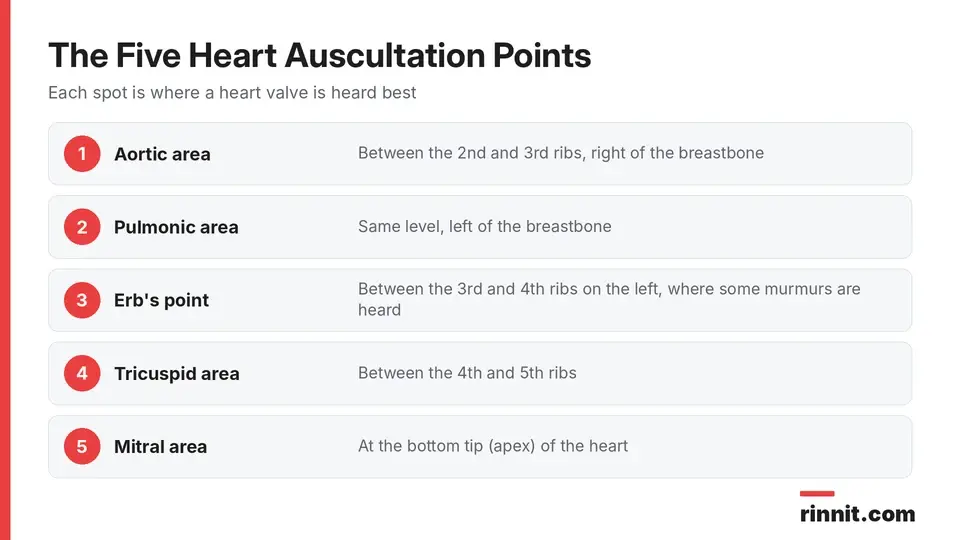
Each point lines up with sounds from a different heart valve.
Each point sits over the area where a particular heart valve is heard best: [1]
Aortic area: between the second and third ribs, right of the breastbone
Pulmonic area: the same level, left of the breastbone
Erb's point: between the third and fourth ribs on the left, where some murmurs are heard
Tricuspid area: between the fourth and fifth ribs
Mitral area: at the bottom tip (apex) of the heart
What do auscultation results mean?
Heart sounds
Normal heart sounds have a steady, two-beat "lub-dub" rhythm. The first sound (S1, "lub") and the second (S2, "dub") are made as your heart valves close. Abnormal findings include: [1]
Murmurs: a swishing sound between beats caused by turbulent blood flow across the valves; some murmurs are harmless and some signal a serious heart condition
Pericardial friction rub: a high-pitched, squeaky sound like two pieces of leather rubbing, which can point to pericarditis
Abnormal heartbeats: an irregular rhythm such as atrial fibrillation, a fast rate of 100 beats per minute or more (tachycardia) or a slow rate of 60 or fewer (bradycardia)
Lung sounds

Common abnormal lung sounds and what they may point to.
Normal lung sounds are soft and hollow and are heard as you breathe in. Abnormal sounds include: [1]
Sound | What it sounds like | Possible causes |
Stridor | Loud, high-pitched | Upper airway blockage, such as epiglottitis, anaphylaxis or vocal cord dysfunction |
Wheeze | High-pitched hissing | Narrowed airways, as in asthma, COPD or pulmonary edema |
Rhonchi | Like snoring | Airway blockage from pneumonia, COPD or cystic fibrosis |
Crackles (rales) | Like two strands of hair rubbing together | Fluid in the airways from pneumonia or heart failure |
Pleural rub | Like sandpaper rubbing | Irritated tissue around the lungs, as in pleurisy, pneumothorax or pleural effusion |
Squawk | A short squeak, between a wheeze and a crackle | Pulmonary fibrosis, bronchiolitis or pneumonia |
Sources: [1]
Bowel sounds
Normal bowel sounds are low-pitched gurgles heard every 5 to 10 seconds as your intestines move food along. Possible abnormal findings: [1]
No sounds for more than two minutes: may mean an ileus, when the intestines temporarily stop working
Reduced sounds: slowed activity or constipation
Increased sounds: faster activity or diarrhea
Grumbling or high-pitched sounds: may signal a bowel obstruction
What is the triangle of auscultation?
The triangle of auscultation is a small area on your back bordered by the trapezius, the latissimus dorsi and the shoulder blade. When you cross your arms and lean forward, the muscles thin out over this spot, so your provider can hear your lungs more clearly there. [1]
Is auscultation safe?
Yes. Auscultation is non-invasive and painless; the stethoscope simply rests on your skin. If your provider hears something unusual, it gives them clues for further testing, so ask about what they found and what comes next. [1]
Conclusion
Auscultation is a quick, safe way for your provider to check your heart, lungs, blood vessels and digestion with nothing more than a stethoscope. Normal sounds are reassuring, and unusual ones help guide which tests you need next.
If your provider hears a murmur, wheeze or other unusual sound, ask what it might mean and whether you need an echocardiogram, imaging or other follow-up.
Keep learning: Read our heart murmurs guide or our pneumonia guide. For more background, see MedlinePlus on auscultation.
FAQ
What is auscultation?
It is listening to the sounds of your heart, lungs, arteries and abdomen with a stethoscope during a physical exam.
Why do doctors use auscultation?
To check your circulatory, respiratory and digestive systems and to help confirm or rule out medical conditions.
Does auscultation hurt?
No. It is a safe, non-invasive exam; the stethoscope just rests on your skin.
What does a normal heartbeat sound like?
A steady two-beat "lub-dub" rhythm, called S1 and S2, made as the heart valves close.
What is a heart murmur on auscultation?
A swishing sound between heartbeats caused by turbulent blood flow across the heart valves.
What abnormal lung sounds can a doctor hear?
Stridor, wheezes, rhonchi, crackles, a pleural rub or a squawk, each linked to different airway or lung problems.
What do crackles in the lungs mean?
Crackles can come from fluid in the airways, as in pneumonia or congestive heart failure.
What do absent bowel sounds mean?
No bowel sounds for more than two minutes may mean an ileus, when the intestines temporarily stop working.
What are the five heart auscultation points?
The aortic area, pulmonic area, Erb's point, tricuspid area and mitral area.
What is the triangle of auscultation?
A small area on the back where muscles thin out so lung sounds can be heard more clearly.
Related Reading
Heart Murmurs: What They Mean, When They're Harmless, and How They're Treated explains the swishing sound a provider may hear on heart auscultation.
Pericarditis: Symptoms, Causes & Treatment — Why Sharp Chest Pain Needs a Prompt Check covers the condition behind a pericardial friction rub.
Pneumonia: Complete Guide to Symptoms, Causes, and Treatment describes an infection that can cause crackles and rhonchi.
Asthma: Symptoms, Causes, Triggers, Diagnosis & Treatment — Complete Guide explains a common cause of wheezing.
What Is Chronic Obstructive Pulmonary Disease (COPD)? Symptoms, Causes, Stages, and Treatment covers a lung disease that can cause wheezes and rhonchi.
Heart Failure: Symptoms, Causes & Treatment Guide explains a heart condition that can cause crackles from fluid in the lungs.
References
Cleveland Clinic. Auscultation: Definition, Purpose & Procedure. Cleveland Clinic. Last updated October 7, 2024. https://my.clevelandclinic.org/health/diagnostics/23080-auscultation
Martindale JL, Wakai A, Collins SP, et al. Diagnosing Acute Heart Failure in the Emergency Department: A Systematic Review and Meta-analysis. Acad Emerg Med. 2016;23(3):223-242. PMID 26910112. https://doi.org/10.1111/acem.12878
Draper J, Subbiah S, Bailey R, et al. Murmur clinic: validation of a new model for detecting heart valve disease. Heart. 2019;105(1):56-59. PMID 30049836. https://doi.org/10.1136/heartjnl-2018-313393
Park JS, Park SY, Moon JW, et al. Artificial Intelligence Models for Pediatric Lung Sound Analysis: Systematic Review and Meta-Analysis. J Med Internet Res. 2025;27:e66491. PMID 40249944. https://doi.org/10.2196/66491
Medical disclaimer
This article is for general education only and does not replace advice from a qualified healthcare professional. Talk to your doctor about your own diagnosis and treatment, and seek emergency care right away for severe or sudden symptoms.



Comments