Understanding the Mullerian Duct: Foundations of Reproductive Anatomy

Updated: 3 days ago
Medically reviewed by Dr. Baraa Alnahhal, MD | Last reviewed: October 7, 2026

Quick Answer: What is the Mullerian duct?
The Mullerian duct, or paramesonephric duct, is a paired fetal structure that develops into the female internal reproductive organs [1]. Without anti-Mullerian hormone, the ducts fuse to form the fallopian tubes, uterus, cervix and upper vagina [1] [3], roughly between the third and twelfth weeks of gestation. When the hormone is present, as in male fetuses, the ducts regress [3].
What the research says
Pregnancy is possible with a Mullerian anomaly, but it is more often high-risk. A systematic review and meta-analysis of 47 studies found that women with congenital uterine anomalies had higher odds of preterm birth (OR 3.89, 95% CI 3.11–4.88), fetal malpresentation (OR 11.11, 95% CI 5.74–21.49) and cesarean birth (OR 4.52, 95% CI 2.19–9.31) than women without anomalies (Panagiotopoulos et al., Obstetrics & Gynecology, 2022) [4].
The type of anomaly changes the risk. A systematic review of 9 studies (3,805 women) found that septate and subseptate uteri (canalization defects) were linked to lower clinical pregnancy rates (RR 0.86, 95% CI 0.77–0.96) and more first-trimester miscarriage (RR 2.89, 95% CI 2.02–4.14), while unicornuate, bicornuate and didelphys uteri (unification defects) did not reduce fertility but did raise the rate of preterm birth (RR 2.97, 95% CI 2.08–4.23) (Chan et al., Ultrasound in Obstetrics & Gynecology, 2011) [5].
A 2026 meta-analysis of more than 3.3 million pregnancies points the same way. Across 30 studies, congenital uterine anomalies were associated with miscarriage (OR 1.45, 95% CI 1.13–1.86) and preterm birth before 37 weeks (OR 3.74, 95% CI 1.99–7.02), and the authors concluded that these pregnancies should be treated as high risk (Fahmo et al., Maternal Health, Neonatology and Perinatology, 2026) [6].
Overview of the Mullerian Duct
The development of the Mullerian duct is a foundational stage in human growth. These structures begin as simple tubes and undergo complex transformations to create the internal reproductive anatomy. Understanding this process is essential for recognizing how structural variations occur and how they might influence health from puberty through adulthood.
The Developmental Timeline of Mullerian Ducts
The formation of the Mullerian ducts begins remarkably early in the life of an embryo. Cleveland Clinic reports that the ducts start developing on top of the temporary mesonephric kidneys about three to four weeks into gestation [1]. As the ducts grow, cells develop that form their different layers. Epithelial and mesenchymal cells provide the lining and the stem-cell potential needed for organ formation, and the layers thicken and grow downward into a long, finger-like projection [1].
Between eight and twelve weeks into gestation, the tips of the Mullerian ducts fuse and the first stages of the internal sex organs begin to form [1], including the uterus and the upper vaginal canal. While this is happening, the presence or absence of specific hormones decides the final outcome. In female development, the lack of anti-Mullerian hormone allows the ducts to reach their full potential [1], while in male development, the hormone triggers their regression [3]. Exact week numbers differ slightly between references because pregnancy weeks can be counted from conception or from the last menstrual period.
Developmental Stage | Approximate Timing | Primary Activity |
Early Formation | About 3–4 weeks into gestation | Ducts begin developing on the temporary mesonephric kidneys [1]. |
Cell Layering | Follows early formation | Epithelial and mesenchymal cells form distinct layers that thicken and grow downward into long, finger-like projections [1]. |
Fusion and Organ Formation | 8–12 weeks into gestation | Duct tips fuse and the first structures of the internal sex organs begin to form [1]. |
Hormonal Fork | During sexual differentiation | Without AMH the ducts keep developing into female organs; with AMH they regress [3]. |
Structural Components and Anatomy
The Mullerian ducts are composed of specialized cells that support their rapid growth and eventual transformation. Epithelial cells form the lining of the ducts, protecting them and helping absorb and release important substances [1]. Mesenchymal cells are stem cells [1], so they can grow into the many different cell types needed for the tissues of the reproductive system.
The ducts develop alongside the Wolffian (mesonephric) ducts on the temporary kidneys of the embryo, which are eventually replaced by fully functioning kidneys [1]. This close relationship between the reproductive and urinary systems in early life explains why structural differences in the Mullerian ducts are sometimes accompanied by variations in kidney development, and why doctors may check the kidneys when a Mullerian anomaly is found [3].
Common Mullerian Duct Anomalies
When the standard process of development or fusion is interrupted, it results in what medical professionals call Mullerian duct anomalies. These variations are rare, appearing in approximately 0.1% to 3.0% of live births [1] [3]. Because these structures are internal, many individuals are unaware of these differences until they reach puberty or attempt to conceive.
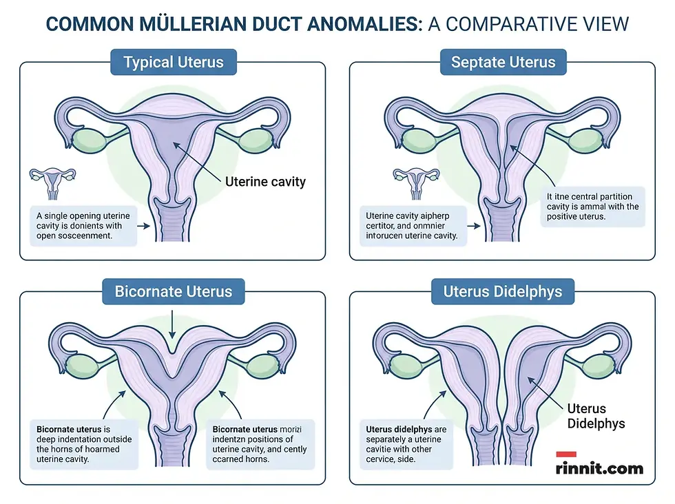
Anomalies can range from the complete absence of certain organs to variations in the shape and structure of the uterus. For instance, Mullerian agenesis occurs when the uterus and/or vagina does not develop fully [1]. Other variations involve the failure of the ducts to fuse or to reabsorb correctly, leading to conditions like a septate, bicornate or didelphys uterus. These structural differences are congenital, meaning they are present from birth, and there is nothing a person can do to prevent them [1].
Type of Anomaly | Description | Potential Impact |
Mullerian Agenesis | Typically no periods; carrying a pregnancy is not possible without medical help [2]. | |
Uterus Didelphys | A double uterus with two separate uterine cavities, typically each with its own cervix. | Higher risk of pregnancy complications [3]; specialized care is often needed. |
Septate Uterus | A band of tissue divides the uterine cavity into two parts. | Increased risk of miscarriage and preterm birth [3]. |
Bicornate Uterus | The ducts did not fully fuse [3], leaving a deep indentation at the top of the uterus. | Pregnancy outcomes vary; some studies link it to a higher risk of complications. |
Persistent Mullerian Duct Syndrome | A rare genetic condition in which biological males also develop a uterus and fallopian tubes [1]. | Undescended testicles; inguinal hernias [1]. |
Recognizing Signs and Seeking Care
The symptoms associated with Mullerian duct variations often become apparent during major reproductive milestones. For many, the first sign is a disruption in the typical menstrual cycle, such as painful or prolonged periods or the total absence of menstruation during puberty [1]. In other cases, individuals may experience pain during sex or when inserting a tampon [1]. Female infertility and kidney problems can also be signs [1], and males with Persistent Mullerian Duct Syndrome may have undescended testicles or inguinal hernias [1].
If a structural difference is suspected, healthcare providers can offer guidance on the available management and treatment options. Diagnosis usually starts with a physical exam and a pelvic ultrasound, and MRI or other imaging may be used for a more detailed view of the internal anatomy [3]. A first period that has not arrived by about age 16 is a good reason to see a provider [2]. Early identification is key to managing potential complications and supporting long-term wellness.

Conclusion
The Mullerian duct is a testament to the complexity of human development. From its early stages as a simple pair of tubes to its final form as the internal reproductive system, it plays a vital role in anatomical identity. While anomalies are rare, understanding the foundations of this developmental process empowers individuals to navigate their health with confidence and clarity.
Next Steps
If you have concerns about your reproductive health or suspect a structural variation, consult with a qualified healthcare professional. Understanding your unique anatomy is the first step toward personalized care and long-term reproductive wellness.
Frequently Asked Questions
What is the Mullerian duct?
The Mullerian duct is a fetal structure that develops into the female internal reproductive organs, including the uterus and fallopian tubes.
When do these ducts start to form?
Cleveland Clinic reports that they begin developing about three to four weeks into gestation.
What organs do the Mullerian ducts create?
The Mullerian ducts form the fallopian tubes, uterus, cervix, and the upper part of the vagina.
Do all fetuses have these ducts?
Yes, all fetuses have Mullerian ducts in the early stages of development.
What determines if the ducts remain or disappear?
The presence of anti-Mullerian hormone (AMH) in male fetuses causes the ducts to regress, while its absence in female fetuses allows them to develop.
What is a Mullerian duct anomaly?
A Mullerian duct anomaly is a structural difference that occurs when the ducts do not develop or fuse as expected.
How common are these anomalies?
Mullerian duct anomalies are rare, occurring in approximately 0.1% to 3.0% of live births.
What are the symptoms of a structural variation?
Symptoms can include painful or prolonged periods, no periods, pain with sex or when inserting a tampon, and difficulty with conception. Some people have no symptoms at all.
Can these issues affect fertility?
Yes, some anomalies can lead to infertility or to complications during pregnancy. The effect depends on the type of anomaly.
Is MRKH syndrome related to the Mullerian duct?
Yes. MRKH syndrome is the most common form of Mullerian agenesis, in which the uterus and upper vagina are missing or underdeveloped.
What is Persistent Mullerian Duct Syndrome?
Persistent Mullerian Duct Syndrome is a rare genetic condition in which biological males, who have typical male reproductive organs, also develop a uterus and fallopian tubes because anti-Mullerian hormone is not released or does not work properly.
Can these anomalies be prevented?
No. They are congenital conditions that occur during fetal development, and there is nothing a person can do to prevent them.
How are these conditions diagnosed?
Mullerian duct conditions are typically identified through a physical examination and imaging tests, such as ultrasound or MRI, performed by a healthcare provider.
Are there treatments available for anomalies?
Yes, various surgical and non-surgical options exist, depending on the specific type of variation. Your healthcare provider can explain which options fit your situation.
Do Mullerian issues affect hormone levels?
In many Mullerian anomalies the ovaries work normally, so estrogen production and puberty changes such as breast development usually proceed as expected. In MRKH syndrome, for example, the ovaries develop from a different structure and are usually unaffected. Ask your healthcare provider how a specific anomaly may affect you.
Related Reading
Understanding MRKH Syndrome: Symptoms, Types, and Care explains a condition where the uterus and vagina do not fully form.
Bicornuate Uterus: The Heart-Shaped Womb You're Born With — and Why Many People Still Have Healthy Pregnancies covers a heart-shaped uterus and its effects on pregnancy.
Double Uterus (Uterus Didelphys): Symptoms, Causes & Treatment — Complete Guide explains a rare condition where two separate uteruses form.
Vaginal Septum: Symptoms, Types, and Treatment Options describes a wall of tissue that can divide the vagina.
Sources
Cleveland Clinic. Müllerian Ducts. Cleveland Clinic. Last updated July 9, 2026. https://my.clevelandclinic.org/health/body/23956-mullerian-duct
Cleveland Clinic. Mayer-Rokitansky-Küster-Hauser Syndrome. Cleveland Clinic. Last updated May 7, 2026. https://my.clevelandclinic.org/health/diseases/23380-mayer-rokitansky-kuster-hauser-syndrome
Wilson D, Bordoni B. Embryology, Mullerian Ducts (Paramesonephric Ducts). In: StatPearls. Treasure Island (FL): StatPearls Publishing; 2026 Jan-. Updated March 6, 2023. PMID 32491659. https://www.ncbi.nlm.nih.gov/books/NBK557727/
Panagiotopoulos M, Tseke P, Michala L. Obstetric complications in women with congenital uterine anomalies according to the 2013 European Society of Human Reproduction and Embryology and the European Society for Gynaecological Endoscopy classification: a systematic review and meta-analysis. Obstet Gynecol. 2022;139(1):138-148. PMID: 34856567. DOI: 10.1097/AOG.0000000000004627
Chan YY, Jayaprakasan K, Tan A, et al. Reproductive outcomes in women with congenital uterine anomalies: a systematic review. Ultrasound Obstet Gynecol. 2011;38(4):371-382. PMID: 21830244. DOI: 10.1002/uog.10056
Fahmo I, Hakizimana T, Kasujja M, et al. Maternal and fetal outcomes in women with uterine congenital abnormalities: a systematic review and meta-analysis. Matern Health Neonatol Perinatol. 2026;12(1):35. PMID: 42563184. DOI: 10.1186/s40748-026-00283-6
Medical disclaimer
This content is for informational purposes only and does not constitute medical advice. Always seek the guidance of a qualified health provider with any questions regarding a medical condition.



Comments